. 2022 Jan 11;88(1):e0148321.
doi: 10.1128/AEM.01483-21. Epub 2021 Oct 27.

Chronic Release of Tailless Phage Particles from Lactococcus lactis

Affiliations

Chronic Release of Tailless Phage Particles from Lactococcus lactis

Yue Liu et al. Appl Environ Microbiol. .

Abstract

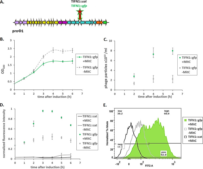
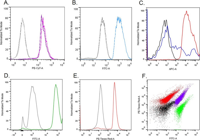
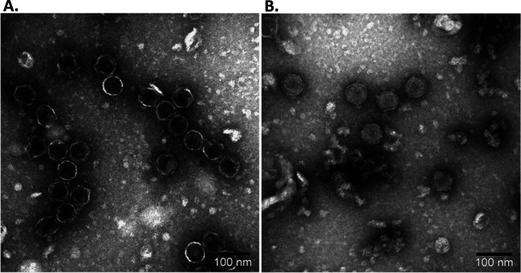
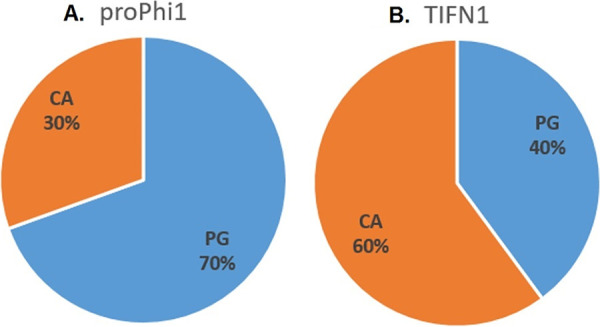
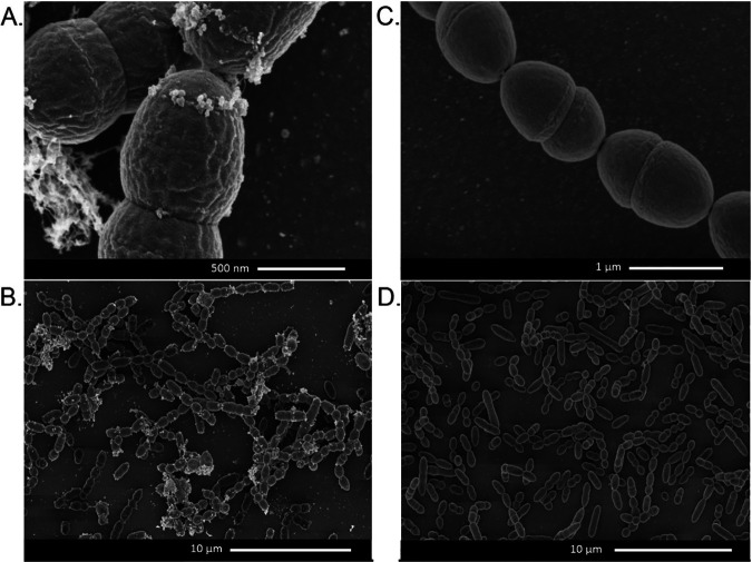
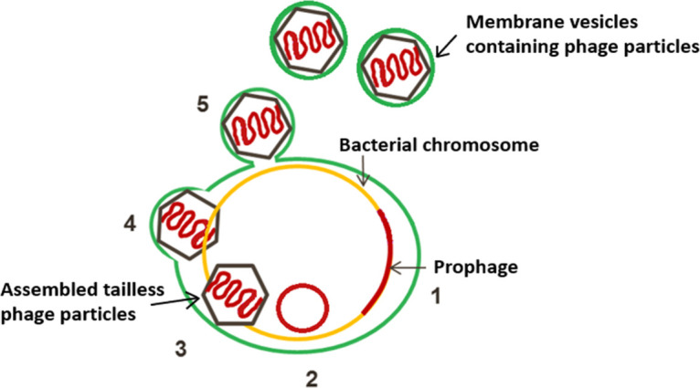
Lactococcus lactis strains residing in the microbial community of a complex dairy starter culture named "Ur" are hosts to prophages belonging to the family Siphoviridae. L. lactis strains (TIFN1 to TIFN7) showed detectable spontaneous phage production and release (109 to 1010 phage particles/ml) and up to 10-fold increases upon prophage induction, while in both cases we observed no obvious cell lysis typically described for the lytic life cycle of Siphoviridae phages. Intrigued by this phenomenon, we investigated the host-phage interaction using strain TIFN1 (harboring prophage proPhi1) as a representative. We confirmed that during the massive phage release, all bacterial cells remain viable. Further, by monitoring phage replication in vivo, using a green fluorescence protein reporter combined with flow cytometry, we demonstrated that the majority of the bacterial population (over 80%) is actively producing phage particles when induced with mitomycin C. The released tailless phage particles were found to be engulfed in lipid membranes, as evidenced by electron microscopy and lipid staining combined with chemical lipid analysis. Based on the collective observations, we propose a model of phage-host interaction in L. lactis TIFN1 where the phage particles are engulfed in membranes upon release, thereby leaving the producing host intact. Moreover, we discuss possible mechanisms of chronic, or nonlytic, release of LAB Siphoviridae phages and its impact on the bacterial host. IMPORTANCE In complex microbial consortia such as fermentation starters, bacteriophages can alter the dynamics and diversity of microbial communities. Bacteriophages infecting Lactococcus lactis are mostly studied for their detrimental impact on industrial dairy fermentation processes. In this study, we describe a novel form of phage-bacterium interaction in an L. lactis strain isolated from a complex dairy starter culture: when the prophages harbored in the L. lactis genome are activated, the phage particles are engulfed in lipid membranes upon release, leaving the producing host intact. Findings from this study provide additional insights into the diverse manners of phage-bacterium interactions and coevolution, which are essential for understanding the population dynamics in complex microbial communities like fermentation starters.

Keywords: Siphoviridae; dairy starter culture; lipid bilayer; membranes; nonlytic phage release.


Figures

FIG 1
FIG 1
Phage labeling and examination of phage replication. (A) Schematic drawing of the prophage genome with marked gfp and cat insertion sites. Arrows represent ORFs and indicate the direction of gene transcription. The number of arrows does not reflect the real ORF numbers but is only a schematic presentation. The insertion was made between two convergent ORFs. Colors in arrows schematically represent different phage gene clusters. (B) Growth response of TIFN1::gfp to MitC treatment. (C) Phage release by TIFN1::gfp during MitC induction. Green symbols represent MitC-treated cultures, and gray symbols represent control cultures without MitC. (D) Dynamics of phage replication (as derived from average cell fluorescence intensity) during MitC induction (green symbols) and in uninduced samples (gray symbols) in reporter strain (TIFN1::gfp) compared to baseline fluorescence of non-gfp cultures (TIFN1::cat, black and gray lines for induced and uninduced conditions, respectively). (E) Fluorescence distribution in the population at 3 h of induction in non-gfp TIFN1::cat (black unfilled), uninduced TIFN1::gfp (gray unfilled), and MitC-induced TIFN1::gfp (green filled) cultures. The statistics in panel E are shown for induced TIFN1::gfp: 82.9% of the population was positive for green fluorescence (pos 82.9), 17.1% was fluorescence negative (neg 17.1), 60.8% was highly fluorescent (high 60.8), and 39.2% was low in fluorescence (low 39.2).
FIG 2
FIG 2
Staining proPhi1 particles with various lipophilic (A, B, C, and F) and DNA binding (D, E, and F) dyes followed by flow cytometry analysis. Gray/black, unstained phage particles. (A) Lipophilic dye 1. (B) Lipophilic dye 2. (C) Lipophilic dye 3. The blue line represents the sample stained after chloroform treatment. (D) DNA dye 1. (E) DNA dye 2. (F) Superimposed dot plot of proPhi1 particle samples with different staining: unstained (black), lipophilic dye 2 (green), DNA dye 2 (red), and double-stained lipophilic dye 2 and DNA dye 2 (purple-blue).
FIG 3
FIG 3
Transmission electron micrograph of proPhi1 with (A) and without (B) chloroform treatment.
FIG 4
FIG 4
Phage and cell lipid composition. Composition of lipids extracted from isolated proPhi1 phage particles (A) and TIFN1 whole-cell-derived protoplast (B). PG, phosphatidyl glycerol; CA, cardiolipin.
FIG 5
FIG 5
Scanning electron micrograph of cells subjected to 6-h MitC treatment. (A and B) TIFN1. (C and D) TI1c.
FIG 6
FIG 6
Schematic presentation of the proposed mechanism (steps 1 to 5) of phage release from Lactococcus lactis TIFN1. Activation of proPhi1 (steps 1 and 2) results in production of tailless Siphoviridae phage particles (step 3), enclosed in lipid membrane derived from the cytoplasmic membrane (green) (step 4), and released from the cells by a budding-like, nonlytic mechanism (step 5).

References

    1. Clokie MRJ, Millard AD, Letarov AV, Heaphy S. 2011. Phages in nature. Bacteriophage 1:31–45. 10.4161/bact.1.1.14942. - DOI - PMC - PubMed
    1. Hatfull GF, Hendrix RW. 2011. Bacteriophages and their genomes. Curr Opin Virol 1:298–303. 10.1016/j.coviro.2011.06.009. - DOI - PMC - PubMed
    1. Ackermann H-W. 2009. Phage classification and characterization. Methods Mol Biol 501:127–140. 10.1007/978-1-60327-164-6_13. - DOI - PubMed
    1. Krupovic M, Prangishvili D, Hendrix RW, Bamford DH. 2011. Genomics of bacterial and archaeal viruses: dynamics within the prokaryotic virosphere. Microbiol Mol Biol Rev 75:610–635. 10.1128/MMBR.00011-11. - DOI - PMC - PubMed
    1. Parmar KM, Gaikwad SL, Dhakephalkar PK, Kothari R, Singh RP. 2017. Intriguing interaction of bacteriophage-host association: an understanding in the era of omics. Front Microbiol 8:559. 10.3389/fmicb.2017.00559. - DOI - PMC - PubMed

Publication types

LinkOut - more resources