. 2021 Nov;64(6):853-863.
doi: 10.3340/jkns.2021.0009. Epub 2021 Oct 28.

Improved Biocompatibility of Intra-Arterial Poly-L-Lactic Acid Stent by Tantalum Ion Implantation : 3-Month Results in a Swine Model

Affiliations

Improved Biocompatibility of Intra-Arterial Poly-L-Lactic Acid Stent by Tantalum Ion Implantation : 3-Month Results in a Swine Model

Kangmin Kim et al. J Korean Neurosurg Soc. 2021 Nov.

Abstract

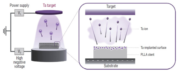
Objective: Biodegradable poly-L-lactic acid (PLLA) with a highly biocompatible surface via tantalum (Ta) ion implantation can be an innovative solution for the problems associated with current biodegradable stents. The purpose of this study is to develop a Ta-implanted PLLA stent for clinical use and to investigate its biological performance capabilities.

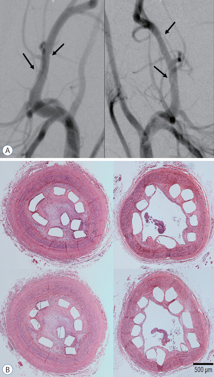
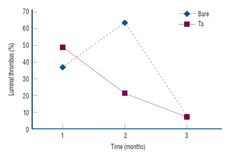
Methods: A series of in vitro and in vivo tests were used to assess the biological performance of bare and Ta-implanted PLLA stents. The re-endothelialization ability and thrombogenicity were examined through in vitro endothelial cell and platelet adhesion tests. An in vivo swine model was used to evaluate the effects of Ta ion implantation on subacute restenosis and thrombosis. Angiographic and histologic evaluations were conducted at one, two and three months post-treatment.

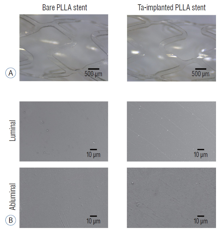
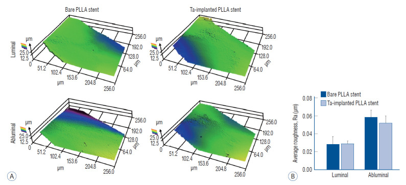
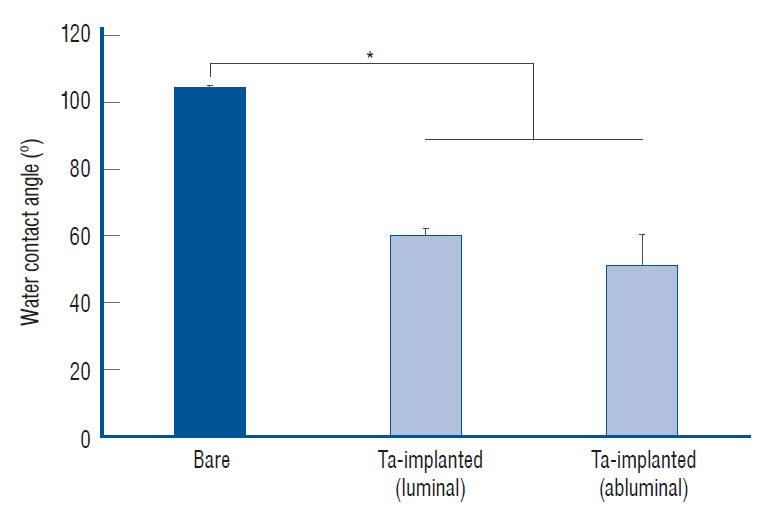
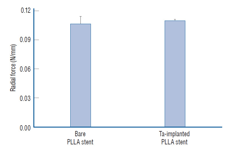
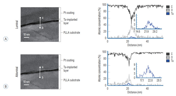
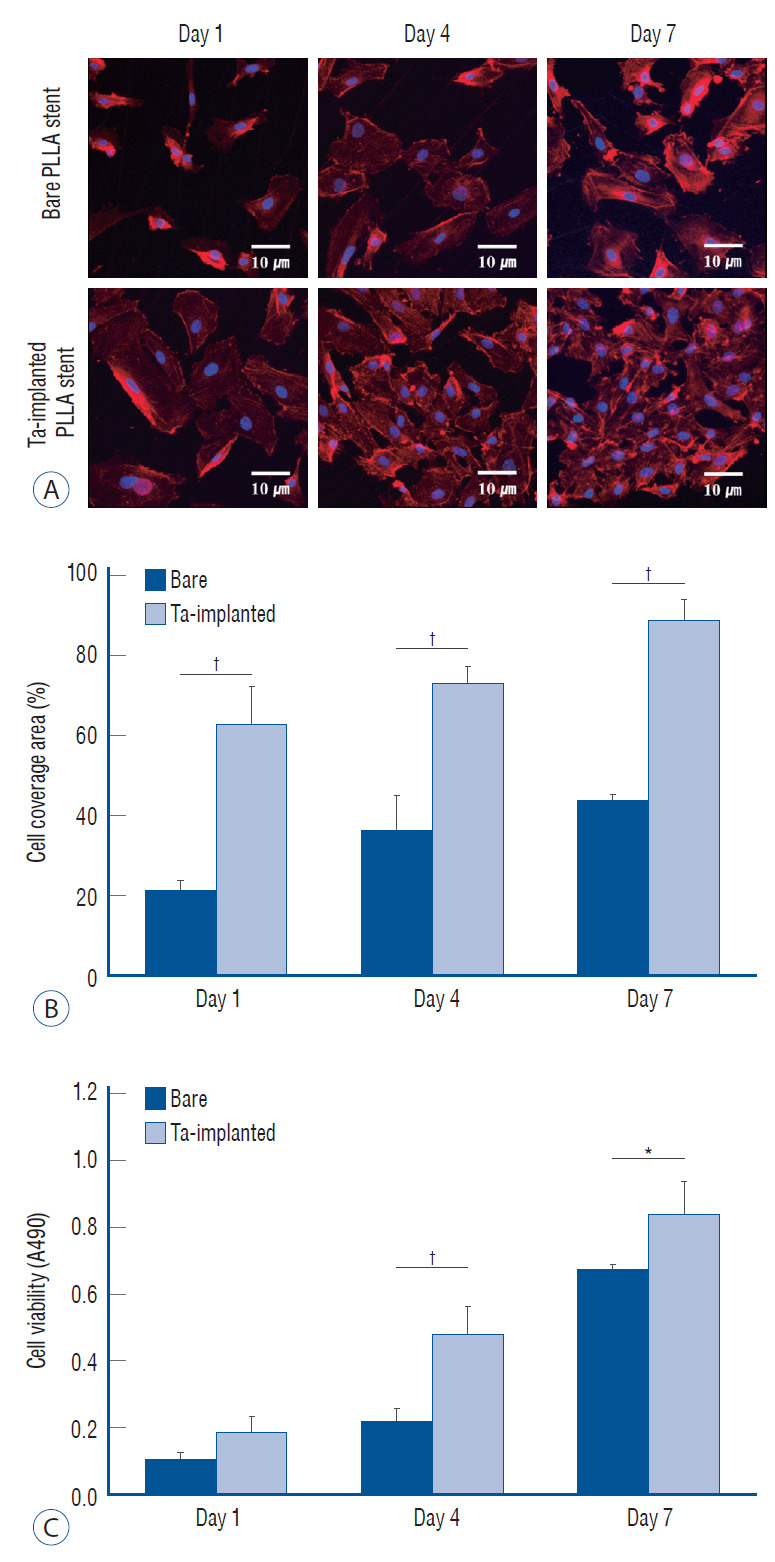
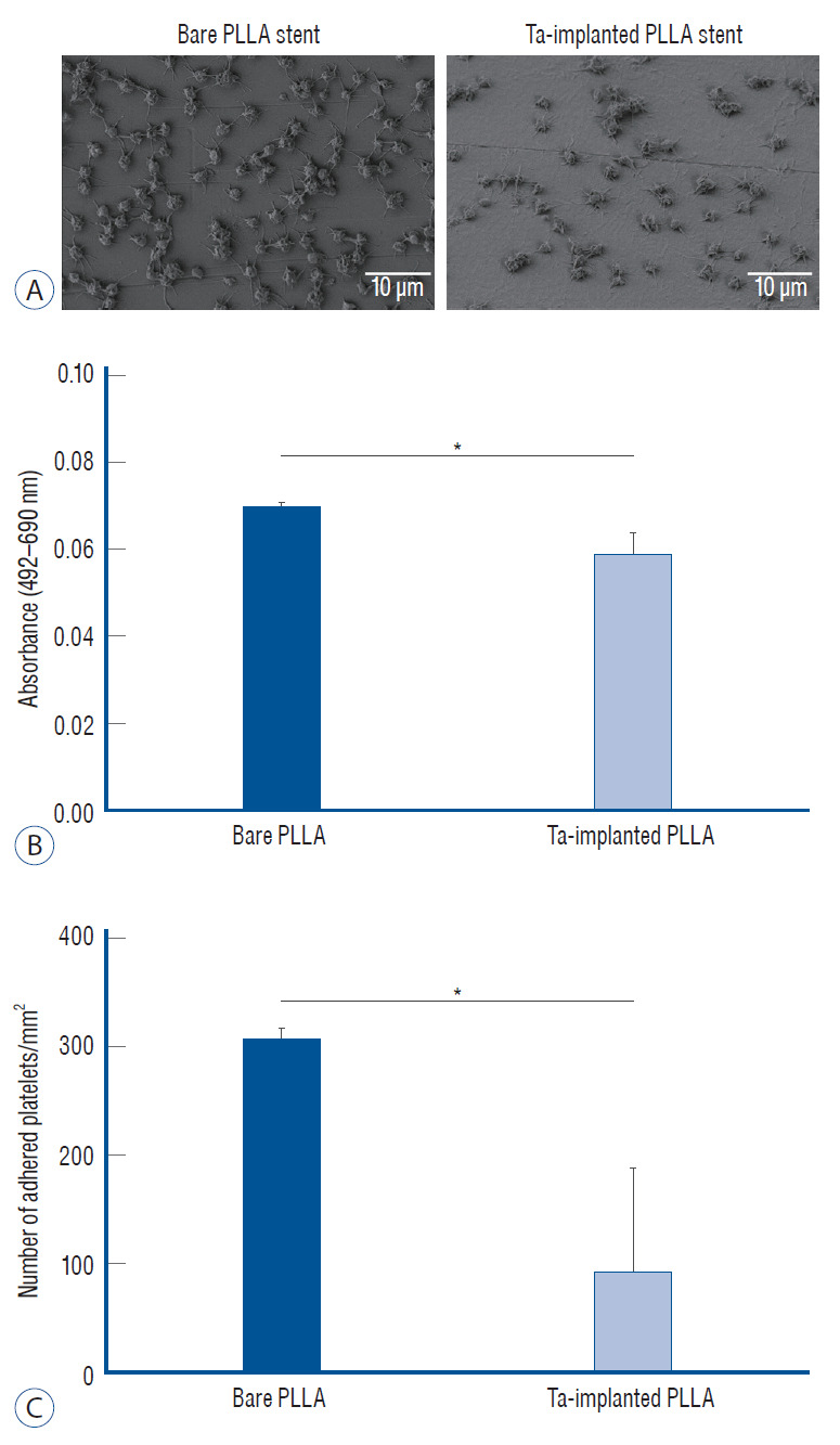
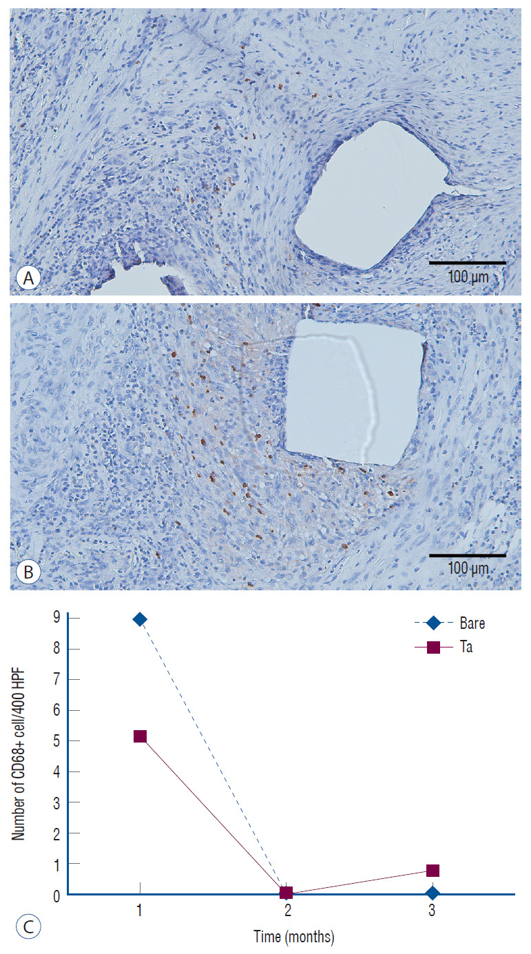
Results: The Ta-implanted PLLA stent was successfully fabricated, exhibiting a smooth surface morphology and modified layer integration. After Ta ion implantation, the surface properties were more favorable for rapid endothelialization and for less platelet attachment compared to the bare PLLA stent. In an in vivo animal test, follow-up angiography showed no evidence of in-stent stenosis in either group. In a microscopic histologic examination, luminal thrombus formation was significantly suppressed in the Ta-implanted PLLA stent group according to the 2-month follow-up assessment (21.2% vs. 63.9%, p=0.005). Cells positive for CD 68, a marker for the monocyte lineage, were less frequently identified around the Ta-implanted PLLA stent in the 1-month follow-up assessments.

Conclusion: The use of a Ta-implanted PLLA stent appears to promote re-endothelialization and anti-thrombogenicity.

Keywords: Absorbable implants; Lactic acid; Pig; Stents; Tantalum.


Conflict of interest statement

CONFLICTS OF INTEREST

No potential conflict of interest relevant to this article was reported.

Figures

Fig. 1.
Fig. 1.
Schematic illustration of the tantalum (Ta) implantation treatment on a poly-L-lactic acid (PLLA) stent. Ta ions were accelerated toward the surface of the PLLA stent, where high negative voltage was applied, forming a Ta-implanted polymer layer.
Fig. 2.
Fig. 2.
Poly-L-lactic acid (PLLA) stent manufactured by laser cutting. The PLLA stent is mounted on a coronary balloon with a gold marker, which is identified in the fluoroscopic image.
Fig. 3.
Fig. 3.
Surface morphology of the stent. Optical (A) and scanning electron microscope (B) images of bare and tantalum (Ta)-implanted poly-L-lactic acid (PLLA) stents. The PLLA stent shows a smooth surface. There is no difference in the surface morphology of the PLLA stent before and after Ta implantation treatment.
Fig. 4.
Fig. 4.
Roughness of the stent. A : Surface topography 3D maps of bare and tantalum (Ta)-implanted poly-L-lactic acid (PLLA) stents. B : There is no significant difference in the calculated average roughness on each surface.
Fig. 5.
Fig. 5.
Water contact angle test. The tantalum (Ta) treatment increased the wettability of the stent. *p<0.05.
Fig. 6.
Fig. 6.
Radial force of the poly-L-lactic acid (PLLA) stent (no significant difference).
Fig. 7.
Fig. 7.
Tantalum layer of the poly-L-lactic acid (PLLA) stent. Representative cross-sectional transmission electron microscope (TEM) image and the TEM/energy-dispersive spectroscopy line profile with c, O, and tantalum (Ta) along the white line from points a to b on the (A) luminal surface and (B) the abluminal surface of the Ta-implanted PLLA stent.
Fig. 8.
Fig. 8.
Endothelialization test. A : confocal laser scanning microscopy images of adhered endothelial cells. Surface coverage of endothelial cells (B) and cell viability (c) on bare and tantalum (Ta)-implanted poly-L-lactic acid (PLLA) surfaces after 1, 4, and 7 days of culturing, respectively. *p<0.05. p<0.01.
Fig. 9.
Fig. 9.
Platelet adhesion test. A : Scanning electron microscope images of the adhered platelet morphology on bare and tantalum-implanted poly-L-lactic acid (PLLA) stents. B : Absorbance difference between 492 nm and 690 nm of a platelet-lysed solution and (c) the number of adhered platelets on each PLLA stent from the lactate dehydrogenase assay. *p<0.05.
Fig. 10.
Fig. 10.
Angiographic and histologic results (×40 magnification) 3 months after stent deployment. A : All parent arteries are patent in the angiographic images without in-stent stenosis (right : tantalum-implanted poly-L-lactic acid [PLLA] stent, left : bare PLLA stent). The black arrows indicate the gold marker of the stent. B : After tissue preparation and hematoxylin and eosin staining with identical specimens, many specimens demonstrate what appears to be an arterial lumen collapse despite the lack of evidence of stenosis in the angiographic images. The discrepancy between the angiographic and histological results may be due to issues that arose during the tissue preparation step with paraffin embedding for the immunohistochemical analysis and stent characteristics such as the small diameter and weak radial force (right : tantalum-implanted PLLA stent, left : bare PLLA stent).
Fig. 11.
Fig. 11.
In vivo thrombogenicity. The tantalum (Ta)-implanted poly-L-lactic acid stent group shows less thrombus formation according to the 2-month follow-up exam (21.2% vs. 63.9%, p=0.005).
Fig. 12.
Fig. 12.
In vivo inflammation. Less of an inflammatory reaction is identified in the tantalum (Ta)-implanted poly-L-lactic acid (PLLA) stent group at 1-month follow-up. The numbers of cD68+ cells/400 HPF are 5.2±7.3 in the Ta-implanted PLLA stent group (A) and 9.0±16.2 in the bare PLLA stent group (B) (×200 magnification). However, these outcomes lack statistical significance (p=0.601). c : The inflammatory response decreases with time.

Similar articles

Cited by

References

    1. Barbarash LS, Bolbasov EN, Antonova LV, Matveeva VG, Velikanova EA, Shesterikov EV, et al. Surface modification of poly-ε-caprolactone electrospun fibrous scaffolds using plasma discharge with sputter deposition of a titanium target. Materials Letters. 2016;171:87–90.
    1. Bledzki AK, Jaszkiewicz A, Scherzer D. Mechanical properties of PLA composites with man-made cellulose and abaca fibres. Compos Part A Appl Sci Manuf. 2009;40:404–412.
    1. Chen JY, Leng YX, Tian XB, Wang LP, Huang N, Chu PK, et al. Antithrombogenic investigation of surface energy and optical bandgap and hemocompatibility mechanism of Ti(Ta+5)O2 thin films. Biomaterials. 2002;23:2545–2552. - PubMed
    1. Collet C, Asano T, Miyazaki Y, Tenekecioglu E, Katagiri Y, Sotomi Y, et al. Late thrombotic events after bioresorbable scaffold implantation: a systematic review and meta-analysis of randomized clinical trials. Eur Heart J. 2017;38:2559–2566. - PubMed
    1. Hanawa T. Metal ion release from metal implants. Mat Sci Eng C-Bio S. 2004;24:745–752.

Grants and funding

LinkOut - more resources