. 2021 Oct;9(10):e003354.
doi: 10.1136/jitc-2021-003354.

4-1BB and optimized CD28 co-stimulation enhances function of human mono-specific and bi-specific third-generation CAR T cells

Affiliations

4-1BB and optimized CD28 co-stimulation enhances function of human mono-specific and bi-specific third-generation CAR T cells

Emiliano Roselli et al. J Immunother Cancer. 2021 Oct.

Abstract

Background: Co-stimulatory signals regulate the expansion, persistence, and function of chimeric antigen receptor (CAR) T cells. Most studies have focused on the co-stimulatory domains CD28 or 4-1BB. CAR T cell persistence is enhanced by 4-1BB co-stimulation leading to nuclear factor kappa B (NF-κB) signaling, while resistance to exhaustion is enhanced by mutations of the CD28 co-stimulatory domain.

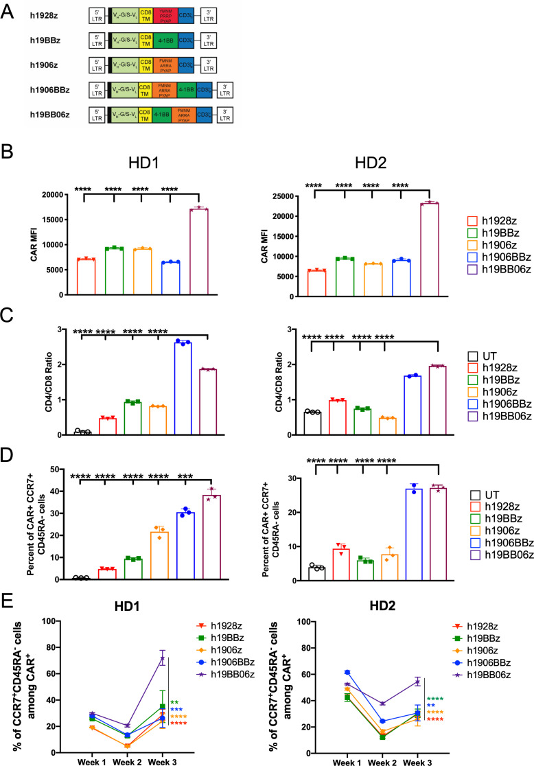
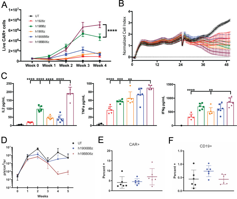
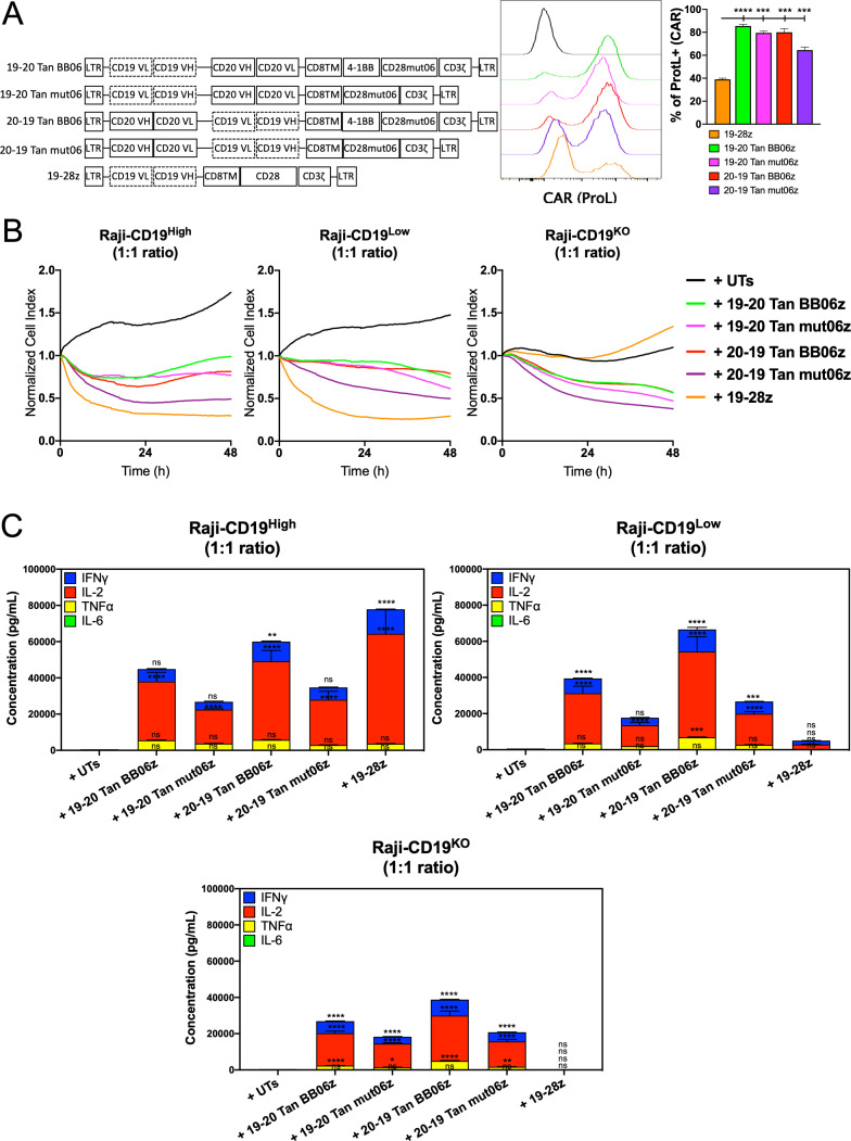
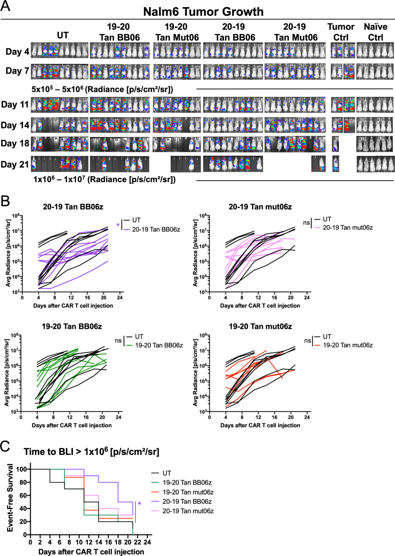
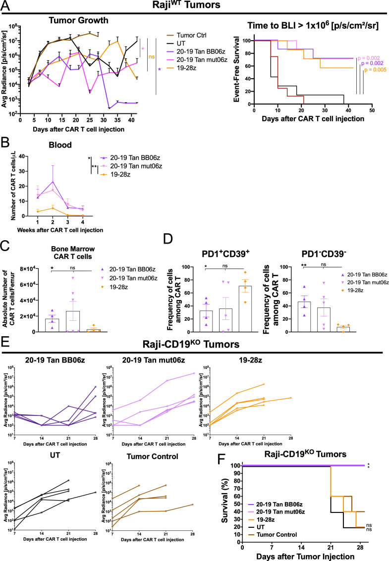
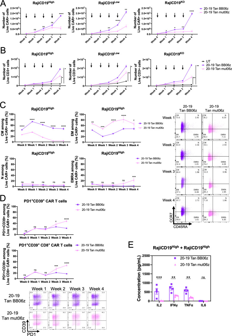
Methods: We hypothesized that a third-generation CAR containing 4-1BB and CD28 with only PYAP signaling motif (mut06) would provide beneficial aspects of both. We designed CD19-specific CAR T cells with either 4-1BB or mut06 together with the combination of both and evaluated their immune-phenotype, cytokine secretion, real-time cytotoxic ability and polyfunctionality against CD19-expressing cells. We analyzed lymphocyte-specific protein tyrosine kinase (LCK) recruitment by the different constructs by immunoblotting. We further determined their ability to control growth of Raji cells in NOD scid gamma (NSG) mice. We also engineered bi-specific CARs against CD20/CD19 combining 4-1BB and mut06 and performed repeated in vitro antigenic stimulation experiments to evaluate their expansion, memory phenotype and phenotypic (PD1+CD39+) and functional exhaustion. Bi-specific CAR T cells were transferred into Raji or Nalm6-bearing mice to study their ability to eradicate CD20/CD19-expressing tumors.

Results: Co-stimulatory domains combining 4-1BB and mut06 confers CAR T cells with an increased central memory phenotype, expansion, and LCK recruitment to the CAR. This enhanced function was dependent on the positioning of the two co-stimulatory domains. A bi-specific CAR targeting CD20/CD19, incorporating 4-1BB and mut06 co-stimulation, showed enhanced antigen-dependent in vitro expansion with lower exhaustion-associated markers. Bi-specific CAR T cells exhibited improved in vivo antitumor activity with increased persistence and decreased exhaustion.

Conclusion: These results demonstrate that co-stimulation combining 4-1BB with an optimized form of CD28 is a valid approach to optimize CAR T cell function. Cells with both mono-specific and bi-specific versions of this design showed enhanced in vitro and in vivo features such as expansion, persistence and resistance to exhaustion. Our observations validate the approach and justify clinical studies to test the efficacy and safety of this CAR in patients.

Keywords: cell engineering; chimeric antigen; immunotherapy; receptors.


Conflict of interest statement

Competing interests: The mut06 CAR has been patented by Moffitt Cancer Center and licensed to Atara Biotherapeutics. MLD has received licensing fees and research support from Atara Biotherapeutics.

Figures

Figure 1
Figure 1
Multiple co-stimulatory domains enhance chimeric antigen receptor (CAR) T cell memory phenotype. (A) Schematic of CAR constructs. All include 5' long terminal repeat (LTR), CD8 signal peptide (black bar), single-chain variable fragment with a variable heavy chain connected with glycine/serine linker to a variable light chain (VH-G/S-VL), CD8α transmembrane and hinge domain, co-stimulatory, and CD3ζ endodomain, and 3' LTR. (B–D) CAR T cells were stimulated with irradiated 3T3-hCD19 target cells at a 10:1 E:T ratio for 24 hours. Cells were then collected, and phenotype determined by flow cytometry. (B) Surface CAR expression on live CD3+CAR+ cells measured by median fluorescent intensity within protein L+ cells. (C) CD4/CD8 ratio on live CD3+CAR+ T cells. (D) Central memory phenotype (CCR7+CD45RA+) among CD3+CAR+ cells at 24 hours. (E) Central memory phenotype (CCR7+CD45RA+) among CD3+CAR+ cells at weeks 1, 2 and 3 after repeated weekly antigen stimulation with CD19+ target cells. Data are shown from two healthy donors (HD). Data are shown as mean±SD. One-way analysis of variance (ANOVA) was performed with Dunnet’s multiple comparison test against h19BB06z for (B–D). Two-way ANOVA was performed with Dunnet’s multiple comparison test against h19BB06z for (E).**p<0.01, ***p<0.001, ****p<0.0001.
Figure 2
Figure 2
Increased phospho-lymphocyte-specific protein tyrosine kinase (pLCK) enhances in vitro function of h19BB06z chimeric antigen receptor (CAR) T cells. (A) CAR T cells were stimulated with irradiated 3T3-hCD19 cells at a 10:1 E:T ratio. After 24 hours, supernatants were harvested, and cytokines were measured with ELLA. Data are shown from three healthy donors (HDs). (B) A single-cell measure of polyfunctionality (top) and Polyfunctional Strength Index (PSI) (bottom) of CAR T cells stimulated for 4 hours with CD19+ target cells. (C) CAR T cells were co-cultured with irradiated 3T3-hCD19 at indicated E:T ratios. The xCELLigence real-time cell analysis (RTCA) system monitored real-time cytotoxicity. (D–E) CAR T cells were stimulated with irradiated 3T3-hCD19 cells at a 10:1 E:T ratio for 24 hours. Cells were lysed and either total lysate (D) or CAR bound and unbound fractions (E) were western blotted. Data are representative of two HDs (B–E). Data are shown as mean±SD. One-way analysis of variance (ANOVA) was performed with Dunnet’s multiple comparison test against h19BB06z for (A). *p<0.05, **p<0.01, ***p<0.001, ****p<0.0001.
Figure 3
Figure 3
h19BB06z chimeric antigen receptor (CAR) T cells have greater proliferation, cytotoxicity, and IL-2 production after repeated antigen stimulations with improved in vivo antitumor activity. (A) 5×10⁵ CAR T cells were stimulated with 1×10⁵ irradiated 3T3-hCD19 cells. After 1 week, half of the cells were enumerated by flow cytometry and the other half was re-stimulated with 1×10⁵ fresh irradiated 3T3-hCD19 cells. This was repeated for a total of 4 weeks. (B–C) After 4 weeks of re-stimulation the same number of CAR T cells for each group was co-cultured with 3T3-hCD19 cells at a 5:1 E:T ratio and either cytotoxicity (B) or cytokine secretion (C) were measured. n=6. Data are representative of 3 (A) or 2 (B–C) healthy donors. Data are shown as mean±SD. (D) NOD scid gamma mice were intravenously injected with 5×10⁵ Raji-GFP/luc cells at week −1 and 2×10⁶ CAR T cells were intravenously injected at week 0. Mice were then measured for bioluminescence imaging (BLI) weekly. Average BLI for each week is shown. (E) Percentage of Live CAR+ cells at week 5. (F) Percentage of live CD19+ cells at week 5. n=6. Data are representative of two independent experiments. Two-way analysis of variance (ANOVA) was performed with Dunnet’s multiple comparison test against h19BB06z for (A). One-way ANOVA was performed with Dunnet’s multiple comparison test against h19BB06z for (C). **p<0.01, ***p<0.001, ****p<0.0001.
Figure 4
Figure 4
CD19/CD20 bi-specific constructs are highly expressed and confer chimeric antigen receptor (CAR) T cells cytotoxicity and cytokine production. (A) Schematic of bi-specific single CAR constructs targeting CD19 and CD20 in a tandem (Tan) configuration. Histograms showing CAR expression. Quantitative analysis of CAR expression (Protein L staining) at day 7 post-transduction compared with 19-28z cells. (B) Real-time cytotoxicity assay (xCelligence) was performed with the different bi-specific CAR T cells and single 19-28z against Raji-CD19High, Raji-CD19Low and Raji-CD19KO target cells at 1:1 E:T ratio. (C) Concentration of interferon gamma (IFN-γ), interleukin (IL)-2, IL-6 and tumor necrosis factor alpha (TNF-α) measured by ELLA in supernatants from co-cultures of bi-specific CAR T cells and single 19-28z with Raji-CD19-High, Raji-CD19Low and Raji-CD19KO after 24 hours compared with untransduced T (UT) cells. All conditions were normalized to the lowest CAR expression using UT cells, reaching same number of CAR T cells and total T cells per group. Representative results of three independent experiments are shown. Data are shown as mean±SD. NS, not significant; *p<0.05, **p<0.01, ***p<0.001, ****p<0.0001. One-way analysis of variance (ANOVA) was performed with Dunnet’s multiple comparison test against UT.
Figure 5
Figure 5
Bi-specific chimeric antigen receptor (CAR) T cells show different in vivo antitumor efficacy. Nalm6-bearing NSG mice were treated with 1x10⁶ CAR T cells 7 days after initial tumor cell injection. (A) Bioluminescent images of each mouse per condition at specified days after T/CAR T cell injection. (B) Tumor burden (average radiance) of each mouse treated with bi-specific CAR T cells and controls. Two-way analysis of variance (ANOVA) was performed with Dunnet’s multiple comparison test against untransduced T cell (UT). Each line represents an individual mouse. (C) Kaplan-Meier analysis of event-free survival defined as time in days to an average luminescence equal or greater than 1x10⁶ p/s/cm2/sr of mice treated with 1x10⁶ T/CAR T cells compared with UT control (n = 8 mice per group). *p<0.05.
Figure 6
Figure 6
CD20-CD19 Tan bi-specific chimeric antigen receptor (CAR) T cells show enhanced in vivo antitumor efficacy and low expression of exhaustion-associated markers. (A) Raji-FFLuc-bearing NSG mice were treated with 1×10⁶ CAR T cells 5 days after initial tumor cell injection. Average tumor burden (average radiance) of mice treated with bi-specific or mono-specific CAR T cells, untransduced T cell (UT) and tumor control. Data are shown as mean±SEM. Two-way analysis of variance (ANOVA) was performed with Dunnet’s multiple comparison test against UT (left). Kaplan-Meier analysis of event-free survival defined as time in days to an average luminescence equal or greater than 1×10⁶ p/s/cm2/sr of mice treated with 1×10⁶ T/CAR T cells compared with UT control (right). *p<0.05. (B) Total number of CAR T cells per μL of blood of tumor-bearing mice treated with the different CAR T cells (each point is 1 mouse). Two-way ANOVA was performed with Dunnet’s multiple comparison test against 19-28z condition. *p<0.05, **p<0.01. (C) Total number of CAR T cells per femur of tumor-bearing mice treated with different CAR T cells (each point is 1 mouse). (D) Expression of PD1 and CD39 among CAR+ cells harvested from the bone marrow of tumor-bearing mice treated with different CAR T cells (n=7 mice per group). Data are shown as mean±SEM. One-way ANOVA was performed with Dunnet’s multiple comparison test against the 19-28z condition. (E) RajiCD19KO-bearing NSG mice were treated with 1×10⁶ CAR T cells 5 days after initial tumor cell injection. Tumor burden (average radiance) of mice treated with bi-specific or mono-specific CAR T cells, UT and tumor control. Each line represents an individual mouse (n=5 mice per group). (F) Kaplan-Meier analysis of survival of mice treated with 1×10⁶ T/CAR T cells compared with tumor only control (n=5 mice per group). P values were determined by a one-sided log-rank Mantel-Cox test. NS, not significant; *p<0.05, **p<0.01. All conditions were normalized to the lowest CAR expression using UT cells, reaching same number of CAR-T cells and total T cells per group. See also online supplemental figure S5.
Figure 7
Figure 7
20-19 Tan BB06z bi-specific chimeric antigen receptor (CAR) T cells show increased expansion, memory phenotype and lower exhaustion-associated markers after repeated antigen stimulation. 5×10⁵ CAR T cells were co-cultured with 5×10⁵ target cells (Raji-CD19KO, Raji-CD19Low and Raji-CD19High). After 1 week half the cells were harvested, enumerated and stained by flow cytometry while the other half were re-stimulated with 5×10⁵ fresh target cells (indicated by arrows). This was repeated for a total of 4 weeks. (A) Number of CAR+ cells at each week. (B) Number of total CD3+ cells each week. Two-way analysis of variance (ANOVA) was performed with Dunnet’s multiple comparison test when comparing to control group (untransduced T cell (UT)) (C) Frequency of central memory (CM—CCR7+CD45RA-), naïve (N—CCR7+CD45RA+), effector memory (EM—CCR7-CD45RA-) and terminally differentiated effector (terminally differentiated effector memory cells re-expressing CD45RA (EMRA)—CCR7-CD45RA+) cells at each week among CAR+ cells. Representative dot-plot for CCR7/CD45RA staining at each week (right). (D) Frequency of PD1+CD39+ cells within total CAR T cells at each week (top) and within CD8+ CAR T cells (bottom). Representative dot-plot for PD1/CD39 staining at each week. (E) After 4 weeks of re-stimulation with Raji-CD19High target cells, the same number of CAR T cells were co-cultured for 24 hours with fresh Raji-CD19High cells at a 1:1 E:T ratio and cytokine secretion was measured. Two-way ANOVA was performed with Sidak’s multiple comparison test when comparing 20–19 Tan mut06z to 20–19 Tan BB06z. Data are shown as mean±SEM. NS, not significant; *p<0.05, **p<0.01, ***p<0.001, ****p<0.0001. Data are representative of three healthy donors. See also online supplemental figure S7.

References

    1. Kowolik CM, Topp MS, Gonzalez S, et al. . Cd28 costimulation provided through a CD19-specific chimeric antigen receptor enhances in vivo persistence and antitumor efficacy of adoptively transferred T cells. Cancer Res 2006;66:10995–1004. 10.1158/0008-5472.CAN-06-0160 - DOI - PubMed
    1. Zhao Z, Condomines M, van der Stegen SJC, et al. . Structural design of engineered costimulation determines tumor rejection kinetics and persistence of CAR T cells. Cancer Cell 2015;28:415–28. 10.1016/j.ccell.2015.09.004 - DOI - PMC - PubMed
    1. Neelapu SS, Locke FL, Bartlett NL, et al. . Axicabtagene Ciloleucel CAR T-cell therapy in refractory large B-cell lymphoma. N Engl J Med Overseas Ed 2017;377:2531–44. 10.1056/NEJMoa1707447 - DOI - PMC - PubMed
    1. Schuster SJ, Svoboda J, Chong EA, et al. . Chimeric antigen receptor T cells in refractory B-cell lymphomas. N Engl J Med 2017;377:2545–54. 10.1056/NEJMoa1708566 - DOI - PMC - PubMed
    1. Philipson BI, O'Connor RS, May MJ, et al. . 4-1BB costimulation promotes CAR T cell survival through noncanonical NF-κB signaling. Sci Signal 2020;13:eaay8248. 10.1126/scisignal.aay8248 - DOI - PMC - PubMed

Publication types

Substances