. 2021 Oct 27;12(1):6197.
doi: 10.1038/s41467-021-26499-y.

A Newcastle disease virus expressing a stabilized spike protein of SARS-CoV-2 induces protective immune responses

Affiliations

A Newcastle disease virus expressing a stabilized spike protein of SARS-CoV-2 induces protective immune responses

Weina Sun et al. Nat Commun. .

Abstract

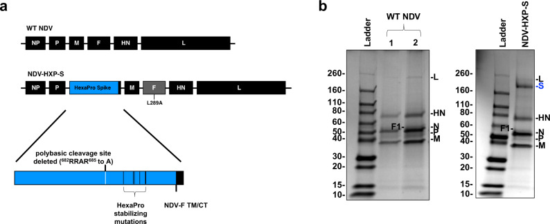
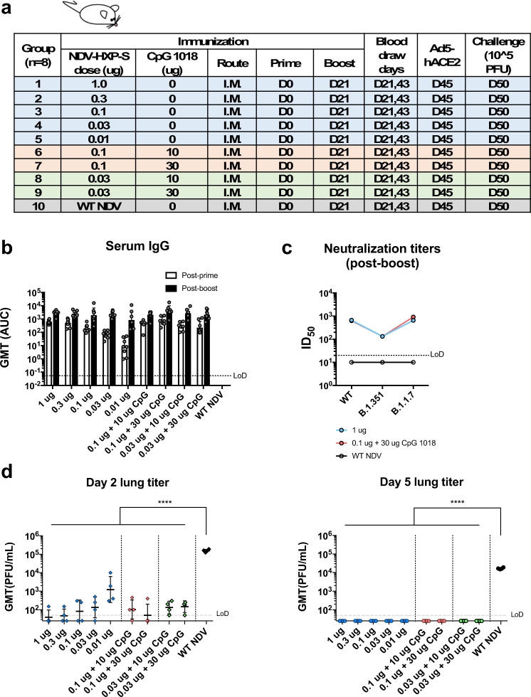
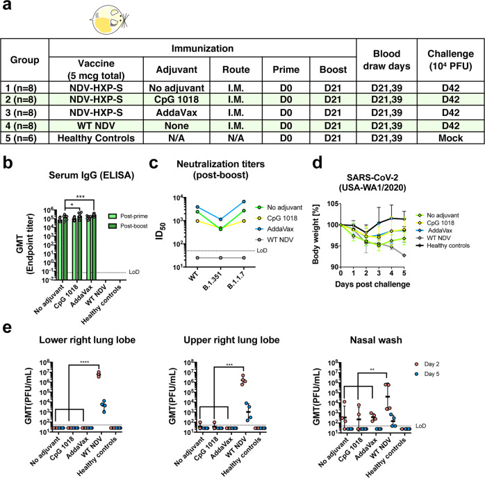
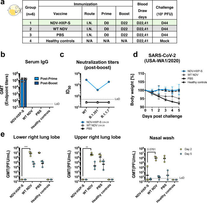
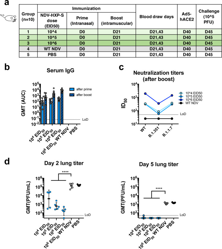
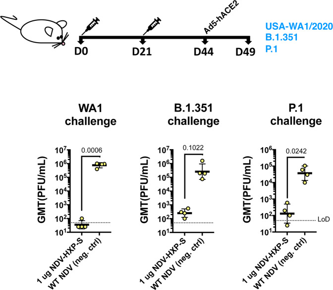
Rapid development of COVID-19 vaccines has helped mitigating SARS-CoV-2 spread, but more equitable allocation of vaccines is necessary to limit the global impact of the COVID-19 pandemic and the emergence of additional variants of concern. We have developed a COVID-19 vaccine candidate based on Newcastle disease virus (NDV) that can be manufactured at high yields in embryonated eggs. Here, we show that the NDV vector expressing an optimized spike antigen (NDV-HXP-S) is a versatile vaccine inducing protective antibody responses. NDV-HXP-S can be administered intramuscularly as inactivated vaccine or intranasally as live vaccine. We show that NDV-HXP-S GMP-produced in Vietnam, Thailand and Brazil is effective in the hamster model. Furthermore, we show that intramuscular vaccination with NDV-HXP-S reduces replication of tested variants of concerns in mice. The immunity conferred by NDV-HXP-S effectively counteracts SARS-CoV-2 infection in mice and hamsters.


Conflict of interest statement

The Icahn School of Medicine at Mount Sinai has filed patent applications entitled “RECOMBINANT NEWCASTLE DISEASE VIRUS EXPRESSING SARS-COV-2 SPIKE PROTEIN AND USES THEREOF” which names P.P., F.K., AG-S., and W.S. as inventors. The AG-S laboratory has received research support from Pfizer, Senhwa Biosciences, Kenall Manufacturing, Avimex, Johnson & Johnson, Dynavax, 7Hills Pharma, Pharmamar, ImmunityBio, Accurius, Merck and Nanocomposix, and AG-S has consulting agreements for the following companies involving cash and/or stock: Vivaldi Biosciences, Contrafect, 7Hills Pharma, Avimex, Vaxalto, Pagoda, Accurius, Esperovax, Farmak, Applied Biological Laboratories, and Pfizer. All other authors declared no competing interests.

Figures

Fig. 1
Fig. 1. Design of the NDV-HXP-S construct.
a Structure and design of the NDV-HXP-S genome. The ectodomain of the spike was connected to the transmembrane domain and cytoplasmic tail (TM/CT) of the F protein (blue: the ectodomain of the spike; black: NDV components; gray: NDV F gene with L289A mutation). The original polybasic cleavage site was removed by mutating RRAR to A. The HexaPro (F817P, A892P, A899P, A942P, K986P, and V987P) stabilizing mutations were introduced. The sequence was codon-optimized for mammalian host expression. b Protein staining of NDV-HXP-S. WT NDV as well as NDV-HXP-S were partially purified from the allantoic fluid through a sucrose cushion and resuspended in PBS. In all, 5 µg (1) and 10 µg (2) of the WT NDV, as well as 10 µg of NDV-HXP-S were resolved on 4–20% SDS–PAGE. The viral proteins were visualized by Coomassie Blue staining (L, S[Blue], HN, N, P, and M). A representative image out of more than three independent experiments is shown.
Fig. 2
Fig. 2. Low doses of inactivated NDV-HXP-S induce a protective antibody response in mice.
a Design of the study. Nine-to-ten-week-old female BALB/c mice were used. Group 1–5 (n = 8) were vaccinated with unadjuvanted NDV-HXP-S at 1 µg, 0.3 µg, 0.1 µg, 0.03 µg, and 0.01 µg per mouse, respectively. Group 6 and 7 (n = 8) were vaccinated with 0.1 µg of NDV-HXP-S with 10 or 30 µg of CpG 1018 per mouse, respectively. Group 8 and 9 (n = 8) were vaccinated with 0.03 µg of NDV-HXP-S with 10 or 30 µg of CpG 1018 per mouse, respectively. Group 10 (n = 8) was vaccinated with 1 µg of WT NDV as the negative control. The vaccine was administered via the intramuscular (I.M.) route at D0 and D21. Blood was collected at D21 and D43. Mice were sensitized with Ad5-hACE2 at D45 and challenged with 105 PFU of the USA-WA1/2020 strain. b Spike-specific serum IgG. Antibodies in post-prime (D21) and post-boost (D43) sera were measured by ELISAs. Geometric mean titer (GMT) represented by the area under the curve (AUC) was graphed, with geometric standard deviation (SD) as error bars. c Neutralizing activity of serum antibodies. Post-boost sera from group 1, group 7, and group 10 were pooled within each group and tested in neutralization assays against the USA-WA1/2020 strain (WT), the B.1.351 variant, and B.1.1.7 variant in technical duplicate. Serum dilutions inhibiting 50% of the infection (ID50) were plotted. (LoD: limit of detection; LoD = 1:20; An ID50 = 1:10 was assigned to negative samples). d Viral load in the lungs. Lungs of a subset of animals (n = 4) from each group were collected on day 2 and day 5 post challenge. The whole lungs were homogenized in 1 mL of PBS. Viral titers were measured by plaque assay on Vero E6 cells and plotted as GMT of PFU/mL (LoD = 50 PFU/mL; a titer of 25 PFU/mL was assigned to negative samples). The error bars represent geometric SD. Statistical difference was analyzed by ordinary one-way ANOVA corrected for Dunnett’s multiple comparisons test (****p < 0.0001). (blue: unadjuvanted groups; red: 0.1 µg adjuvanted with CpG 1018; green: 0.03 µg adjuvanted with CpG 1018).
Fig. 3
Fig. 3. Inactivated NDV-HXP-S induces protective antibody response in hamsters.
a Design of the study. Eighteen-to-twenty-week-old female Golden Syrian hamsters were used. Groups 1–3 (n = 8) were vaccinated with 5 µg of NDV-HXP-S without adjuvants, with CpG 1018 and AddaVax, respectively. Group 4 (n = 8) was vaccinated with 5 µg of WT NDV as the negative control. Group 5 (n = 6) was not vaccinated. The vaccine was administered via the intramuscular (I.M.) route at D0 and D21. Blood was collected at D21 and D39. Group 1–4 were challenged with 104 PFU of USA-WA1/2020 strain at D42. Group 5 was mock-challenged with PBS. b Spike-specific serum IgG. Antibodies in post-prime (D21) and post-boost (D39) sera were measured by ELISAs. GMT endpoint titer was graphed. The error bars represent geometric SD. Statistical difference was analyzed by two-way ANOVA corrected for Dunnett’s multiple comparisons test (*p = 0.0262; ***p = 0.0006). c Neutralizing activity of serum antibodies. Post-boost sera from groups 1–4 were pooled within each group and tested in neutralization assays against USA-WA/2020 strain (WT), B.1.351 variant, and B 1.1.7 variant in technical duplicate (green: no adjuvant; yellow: adjuvanted with CpG 1018; blue: adjuvanted with AddaVax; gray: WT NDV control). Serum dilutions inhibiting 50% of the infection (ID50) were plotted. (LoD = 1:50; an ID50 = 1:25 was assigned to negative samples) d Body weight change of hamsters. Body weights were recorded for 5 days after challenge (green: no adjuvant; yellow: adjuvanted with CpG 1018; blue: adjuvanted with AddaVax; gray: WT NDV control; black: healthy controls). The error bars represent geometric SD. e Viral load in the lungs and nasal washes. The lower right and upper right lung lobes of a subset of animals (n = 4 for groups 1–4; n = 3 for group 5) from each group were collected at day 2 (red) and day 5 (blue) post challenge. Each lung lobe was homogenized in 1 mL PBS. Nasal washes were collected in 0.4 mL of PBS. Viral titers were measured by plaque assay on Vero E6 cells and plotted as GMT of PFU/mL (LoD = 50 PFU/mL; a titer of 25 PFU/mL was assigned to negative samples). The error bars represent geometric SD. Statistical difference was analyzed by two-way ANOVA corrected for Dunnett’s multiple comparisons test (**p = 0.0026 (WT NDV vs. no adjuvant), p = 0.0025 (WT NDV vs. CpG 1018), p = 0.0024 (WT NDV vs. AddaVax); ***p = 0.0001; ****p < 0.0001).
Fig. 4
Fig. 4. GMP lots of inactivated NDV-HXP-S produced by influenza virus vaccine manufacturers induce a protective antibody response in hamsters.
a Design of the study. Nine-to-eleven-week-old female Golden Syrian hamsters were used. Groups 1–6 (n = 8) were vaccinated with 1 µg of spike antigen of inactivated NDV-HXP-S from GPO, IVAC, and Butantan in the absence or presence of CpG 1018. Group 7 (n = 8) was vaccinated with PBS as the negative control. Group 8 (n = 8) was not vaccinated (HC, healthy controls). The vaccine was administered via the intramuscular (I.M.) route at D0 and D21. Blood was collected at D0, D21, and D33. Groups 1–7 were challenged with 104 PFU of the USA-WA1/2020 strain at D35. Group 8 was mock-challenged with PBS. b Spike-specific serum IgG. Antibodies in pre-vaccination (D0, black), post-prime (D21, red), and post-boost (D33, blue) sera were measured by ELISAs (solid bars: unadjuvanted; pattern bars: adjuvanted). GMT endpoint titers were graphed. The error bars represent geometric SD. c Neutralizing activity of serum antibodies. A pseudo-particle neutralization assay was performed by Nexelis to measure neutralization titers of post-boost sera (D33). Human convalescent sera were included in the same assay as the controls (LoD = 1:25; An ID50 = 1:12.5 was assigned to negative samples). d Body weight changes of hamsters. Body weights were monitored for 5 days after the challenge. The error bars represent geometric SD. e Viral load in the lungs. The lower right and upper right Lung lobes of a subset of animals (n = 4) from each group were collected at day 2 and day 5 post challenge. Each lung lobe was homogenized in 1 mL PBS. f Viral load in nasal washes and g nasal turbinates. On day 2 and day 5 post challenge, nasal washes were collected in 0.4 mL of PBS. Nasal turbinates were homogenized in 0.5 mL PBS. Viral titers were measured by plaque assay on Vero E6 cells and plotted as GMT of PFU/mL (LoD = 50 PFU/mL; a titer of 25 PFU/mL was assigned to negative samples). The error bars represent geometric SD. Statistical difference was analyzed by ordinary one-way ANOVA corrected for Dunnett’s multiple comparisons test (****p < 0.0001). cg Green: GPO; red: IVAC; blue: Butantan; gray: PBS: black: healthy controls.
Fig. 5
Fig. 5. Live NDV-HXP-S via the intranasal route induces protective antibody responses in hamsters.
a Design of the study. Eighteen-to-twenty-week-old female Golden Syrian hamsters were used. Group 1 (n = 6) was vaccinated with 106 EID50 of live NDV-HXP-S. Group 2 (n = 6) was vaccinated with 106 EID50 of live WT NDV as the vector-only control. Group 3 (n = 6) was vaccinated with PBS as the negative control. Group 4 (n = 6) were the healthy controls. The vaccine was administered via the intranasal (I.N.) route at D0 and D22. Blood was collected at D22 and D41. Group 1–3 were challenged with 105 PFU of the USA-WA1/2020 strain at D44. Group 4 was mock-challenged with PBS. b Spike-specific serum IgG. Antibodies in post-prime (D22, solid blue bar) and post-boost (D41, pattern blue bar) sera were measured by ELISAs. GMT endpoint titers were graphed. The error bars represent geometric SD. c Neutralizing activity of serum antibodies. Post-boost sera from groups 1–3 were pooled within each group and tested in neutralization assay against USA-WA/2020 strain (WT), B.1.351 variant, and B 1.1.7 variant in technical duplicate. Serum dilutions inhibiting 50% of the infection (ID50) were plotted (LoD = 1:50; An ID50 = 1:25 was assigned to negative samples). d Body weight change of hamsters. Bodyweight was recorded for 5 days after the challenge. The error bars represent geometric SD. (c and d, blue: NDV-HXP-S; gray: WT NDV; black: PBS) e Viral load in the lungs and nasal washes. The lower right and upper right lung lobes of a subset of animals (n = 3) from each group were collected at day 2 (yellow) and day 5 (blue) post challenge. Each lung lobe was homogenized in 1 mL PBS. Nasal washes were collected in 0.4 mL of PBS. Viral titers were measured by plaque assay on Vero E6 cells and plotted as GMT of PFU/mL (LoD = 50 PFU/mL; a titer of 25 PFU/mL was assigned to negative samples). The error bars represent geometric SD. Statistical difference was analyzed by two-way ANOVA corrected for Dunnett’s multiple comparisons test (**p < 0.0017; ***p = 0.0009).
Fig. 6
Fig. 6. Intranasal prime followed by an intramuscular boost of live NDV-HXP-S induces protective antibody responses in mice.
a Design of the study. Seven-to-nine-week-old female BALB/c mice were used. Group 1–3 (n = 10) were vaccinated with 104, 105, and 106 EID50 of live NDV-HXP-S, respectively. Group 4 (n = 10) was vaccinated with 106 EID50 of WT NDV. Group 5 (n = 10) was mock-vaccinated with PBS. The vaccine was administered via the intranasal (I.N.) route at D0 and intramuscular (I.M.) route at D21. Blood was collected at D21 and D43. Mice were sensitized with Ad5-hACE2 at D40 and challenged with 105 PFU of the USA-WA1/2020 strain at D45. b Spike-specific serum IgG. Antibodies in post-prime (D21, solid blue bars) and post-boost (D43, pattern blue bars) sera were measured by ELISAs. GMT AUC was graphed. The error bars represent geometric SD. c Neutralizing activity of serum antibodies. Post-boost sera from groups 1–4 were pooled within each group and tested in neutralization assay against USA-WA/2020 strain (WT), B.1.351 variant, and B 1.1.7 variant in technical duplicate (cyan: 104 EID50; light blue: 105 EID50; blue: 106 EID50; black: WT NDV). Serum dilutions inhibiting 50% of the infection (ID50) were plotted (LoD = 1:50; An ID50 = 1:25 was assigned to negative samples). d Viral load in the lungs. Lungs of a subset of animals (n = 5) from each group were collected at day 2 and day 5 post challenge (blue: NDV-HXP-S; gray: WT NDV; black: PBS). The whole lungs were homogenized in 1 mL PBS. Viral titers were measured by plaque assay on Vero E6 cells and plotted as GMT of PFU/mL (LoD = 50 PFU/mL; a titer of 25 PFU/mL was assigned to negative samples). The error bars represent geometric SD. Statistical difference was analyzed by ordinary one-way ANOVA corrected for Dunnett’s multiple comparisons test (****p < 0.0001).
Fig. 7
Fig. 7. Inactivated NDV-HXP-S induces protective antibody response against the challenge of SARS-CoV-2 variants of concern.
Eight-to-ten-week-old female BALB/c mice were either vaccinated with 1 µg of inactivated NDV-HXP-S (n = 4) or WT NDV (n = 4, negative control). Two immunizations were performed via the intramuscular route at D0 and D21. At D44, mice were treated with Ad5-hACE2. At D49, one-third of mice from each group was challenged with USA-WA1/2020, B.1.351, or P.1 strain. On day 2, lungs were harvested and homogenized in 1 mL PBS. Viral titers were measured by plaque assay on Vero E6 cells and plotted as GMT of PFU/mL (LoD=50 PFU/mL; A titer of 25 PFU/mL was assigned to negative samples). The error bars represent geometric SD. Statistical difference was analyzed by a one-tailed t test. The p values are indicated.

References

    1. Tracking Coranavirus Vaccination Around the World. The New York Times (2021).
    1. Sun, W. et al. A newcastle disease virus (NDV) expressing a membrane-anchored spike as a cost-effective inactivated SARS-CoV-2 vaccine. Vaccines (Basel)8, 10.3390/vaccines8040771 (2020). - PMC - PubMed
    1. Sun W, et al. Newcastle disease virus (NDV) expressing the spike protein of SARS-CoV-2 as a live virus vaccine candidate. EBioMedicine. 2020;62:103132. doi: 10.1016/j.ebiom.2020.103132. - DOI - PMC - PubMed
    1. Hsieh CL, et al. Structure-based design of prefusion-stabilized SARS-CoV-2 spikes. Science. 2020;369:1501–1505. doi: 10.1126/science.abd0826. - DOI - PMC - PubMed
    1. Sparrow E, et al. Global production capacity of seasonal and pandemic influenza vaccines in 2019. Vaccine. 2021;39:512–520. doi: 10.1016/j.vaccine.2020.12.018. - DOI - PMC - PubMed

Publication types

MeSH terms

Substances