Comparative Study
. 2021 Oct 27;11(1):21216.
doi: 10.1038/s41598-021-00642-7.

Predicting and comparing three corrective techniques for sagittal craniosynostosis

Affiliations
Comparative Study

Predicting and comparing three corrective techniques for sagittal craniosynostosis

Connor Cross et al. Sci Rep. .

Abstract

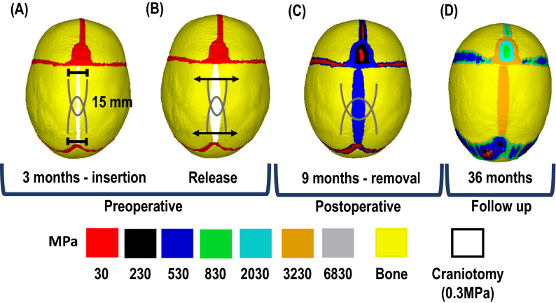
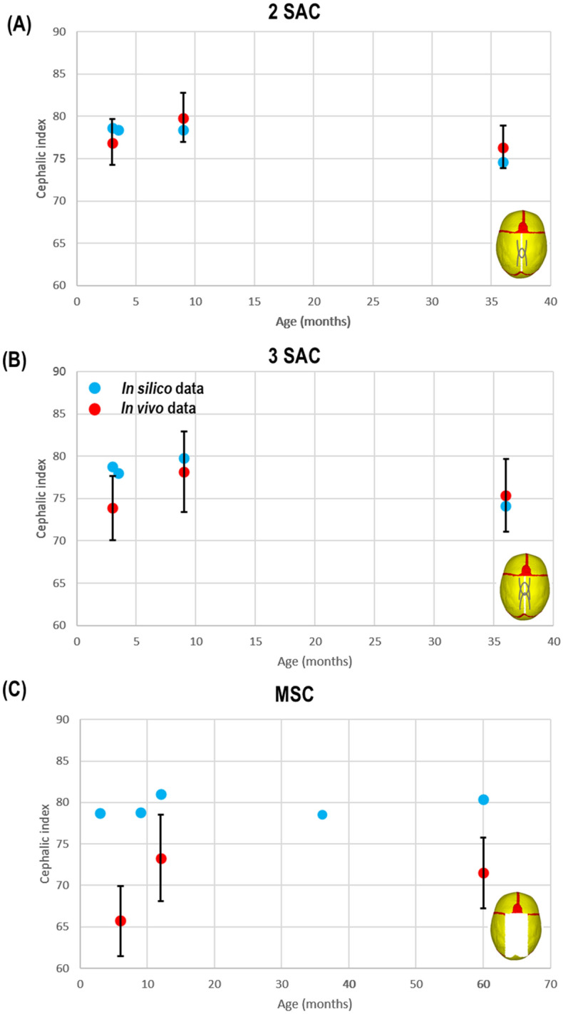
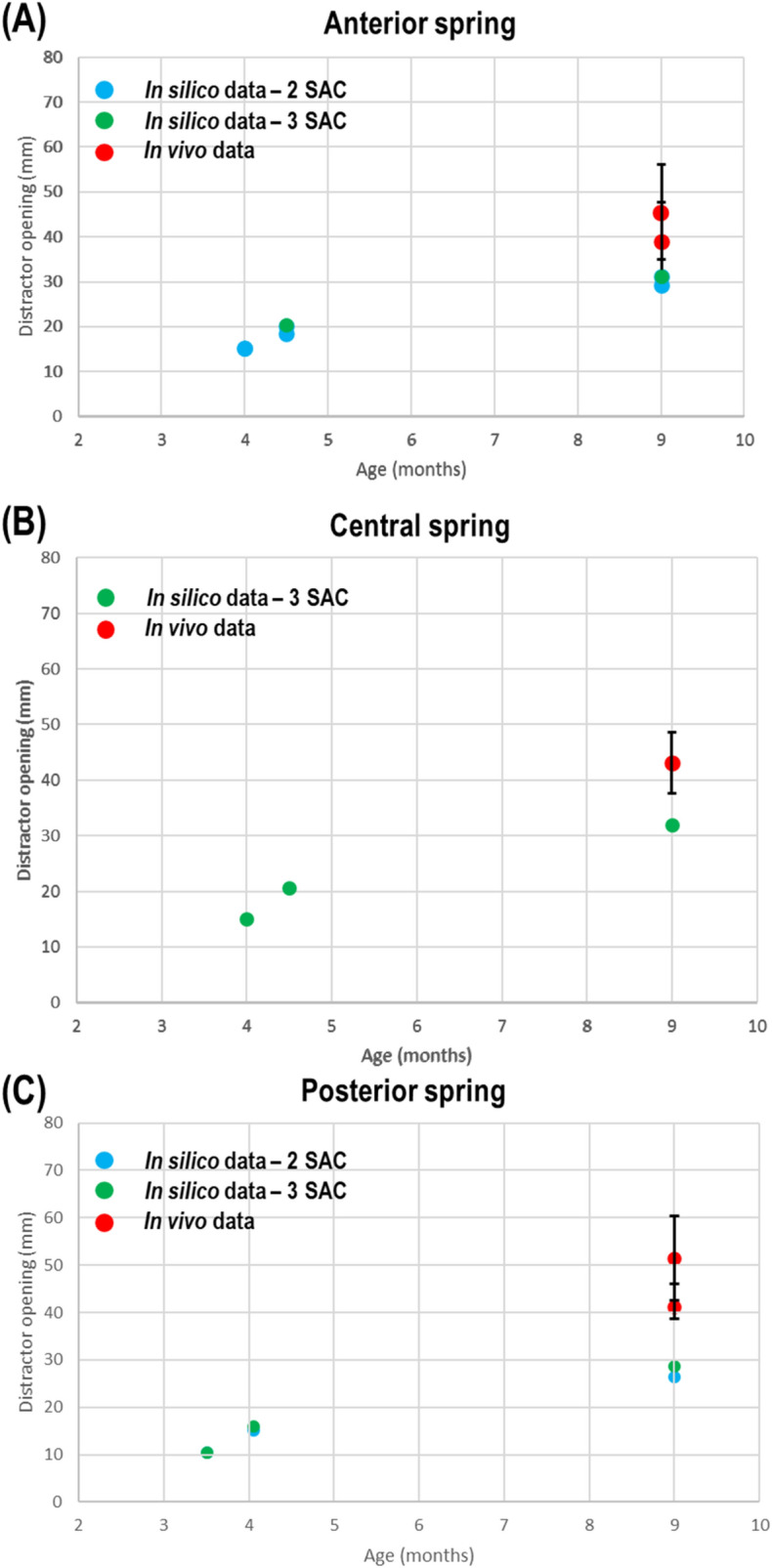
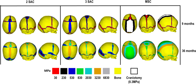
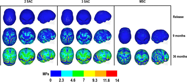
Sagittal synostosis is the most occurring form of craniosynostosis, resulting in calvarial deformation and possible long-term neurocognitive deficits. Several surgical techniques have been developed to correct these issues. Debates as to the most optimal approach are still ongoing. Finite element method is a computational tool that's shown to assist with the management of craniosynostosis. The aim of this study was to compare and predict the outcomes of three reconstruction methods for sagittal craniosynostosis. Here, a generic finite element model was developed based on a patient at 4 months of age and was virtually reconstructed under all three different techniques. Calvarial growth was simulated to predict the skull morphology and the impact of different reconstruction techniques on the brain growth up to 60 months of age. Predicted morphology was then compared with in vivo and literature data. Our results show a promising resemblance to morphological outcomes at follow up. Morphological characteristics between considered techniques were also captured in our predictions. Pressure outcomes across the brain highlight the potential impact that different techniques have on growth. This study lays the foundation for further investigation into additional reconstructive techniques for sagittal synostosis with the long-term vision of optimizing the management of craniosynostosis.


Conflict of interest statement

The authors declare no competing interests.

Figures

Figure 1
Figure 1
Simulation workflow. All techniques were replicated at 4 months of age, when spring insertion (A) and ‘release’ (B) were replicated for SAC. Skull growth, calvarial healing and bone formation at sutures were replicated up to 9 months of age, when springs were removed (C). Skull growth then continued up to 36 months of age [D].
Figure 2
Figure 2
Predicted vs. in vivo cephalic index data with SD. Showing 2 SAC (A), 3 SAC (B) & MSC outcomes (C).
Figure 3
Figure 3
Predicted vs. in vivo spring opening data with SD. Showing anterior (A), central (B) & posterior (C) springs in both SAC techniques. Diagrams show regions where measurements were performed.
Figure 4
Figure 4
2 SAC 3D distance plot at respective ages against mean in vivo CT skull.
Figure 5
Figure 5
3 SAC 3D distance plot at respective ages against mean in vivo CT skull.
Figure 6
Figure 6
Bone formation predictions across sutures/craniotomy across all techniques.
Figure 7
Figure 7
ICV pressure predictions across ICV-bone surface for all techniques.

References

    1. Morriss-Kay GM, Wilkie AOM. Growth of the normal skull vault and its alteration in craniosynostosis: insights from human genetics and experimental studies. J. Anat. 2005;207:637–653. doi: 10.1111/j.1469-7580.2005.00475.x. - DOI - PMC - PubMed
    1. Johnson D, Wilkie AOM. Craniosynostosis. Eur. J. Hum. Genet. 2011;19(4):369–376. doi: 10.1038/ejhg.2010.235. - DOI - PMC - PubMed
    1. Mathijssen IMJ. Guideline for care of patients with the diagnoses of craniosynostosis: working group on craniosynostosis. J. Craniofac. Surg. 2015;26(6):1735–1807. doi: 10.1097/SCS.0000000000002016. - DOI - PMC - PubMed
    1. Kalantar-Hormozi H, Abbaszadeh-Kasbi A, Sharifi G, Davai N, Kalantar-Hormozi A. Incidence of familial craniosynostosis among patients with nonsyndromic craniosynostosis. J. Craniofac. Surg. 2019;30(6):514–517. doi: 10.1097/SCS.0000000000005419. - DOI - PubMed
    1. Rocco FD, Arnaud E, Renier D. Evolution in the frequency of nonsyndromic craniosynostosis. J. Neurosurg. Pediatr. 2009;4(1):21–25. doi: 10.3171/2009.3.PEDS08355. - DOI - PubMed

Publication types