Review
. 2021 Oct;53(10):1539-1546.
doi: 10.1038/s12276-021-00692-x. Epub 2021 Oct 27.

Concept and application of circulating proteasomes

Affiliations
Review

Concept and application of circulating proteasomes

Won Hoon Choi et al. Exp Mol Med. 2021 Oct.

Abstract

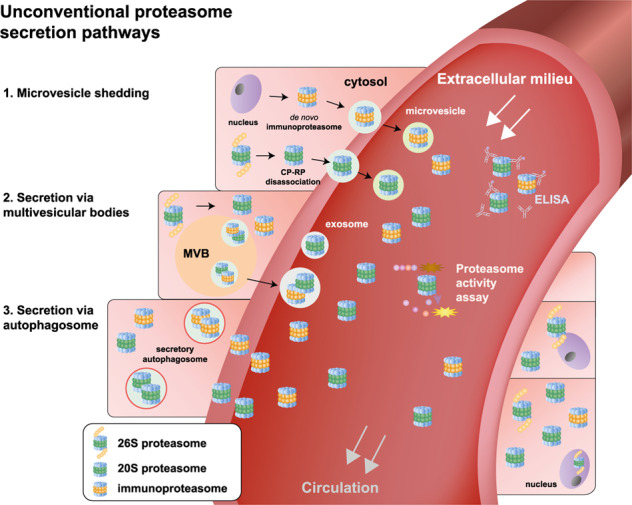
Proteostasis is primarily a function of protein synthesis and degradation. Although the components and processes involved in intracellular proteostasis have been studied extensively, it is apparent that extracellular proteostasis is equitably crucial for the viability of organisms. The 26S proteasome, a unique ATP-dependent proteolytic complex in eukaryotic cells, contributes to the majority of intracellular proteolysis. Accumulating evidence suggests the presence of intact 20S proteasomes in the circulatory system (c-proteasomes), and similar to other plasma proteins, the levels of these c-proteasomes may vary, potentially reflecting specific pathophysiological conditions. Under normal conditions, the concentration of c-proteasomes has been reported to be in the range of ~0.2-2 μg/mL, which is ~2-4-fold lower than that of functional plasma proteins but markedly higher than that of signaling proteins. The characterization of c-proteasomes, such as their origin, structure, role, and clearance, has been delayed mainly due to technical limitations. In this review, we summarize the current perspectives pertaining to c-proteasomes, focusing on the methodology, including our experimental understanding. We believe that once the pathological relevance of c-proteasomes is revealed, these unique components may be utilized in the diagnosis and prognosis of diverse human diseases.


Conflict of interest statement

The authors declare no competing interests.

Figures

Fig. 1
Fig. 1. Activity of circulating proteasomes (c-proteasomes) in human plasma.
a Plasma samples were collected from four individuals (plasma A–D) in EDTA tubes, and their c-proteasome activity (in 20 μL of plasma) was evaluated by monitoring the hydrolysis of the fluorogenic reporter substrates (final concentration of 250 μM in a total of 100 μL reaction), such as suc-LLVY-AMC (for chymotrypsin-like activity), Boc-LRR-AMC (for trypsin-like activity), and Z-LLE-AMC (for caspase-like activity) in the presence or absence of the proteasome inhibitor MG132 (10 μM). These reactions were performed using assay buffer (50 mM Tris-HCl [pH 7.5], 1 mM EDTA, 1 mg/mL BSA, 1 mM ATP, and 1 mM DTT). Sodium dodecyl sulfate (SDS) was not added to the reaction unless otherwise described. The graphs (left) represent the results obtained in three independent experiments, and the mean of the raw fluorescence values (right) at 60 min are plotted with their standard deviations (N = 3). b Human c-proteasome activity was analyzed using suc-LLVY-AMC as the substrate, along with a wide range of protease inhibitors, including aprotinin (trypsin inhibitor), pepstatin A (aspartyl protease inhibitor), and leupeptin (serine/cysteine protease inhibitor). c As in (b), but using different proteasome inhibitors (10 μM MG132, 2 μM bortezomib, 2 μM epoxomicin, or 100 nM carfilzomib). d The plasma samples were preincubated with SDS at the indicated final concentrations for 10 min before initiating the suc-LLVY-AMC hydrolysis reactions. Relative fluorescence values after 30-min reactions were normalized to those obtained in the presence of 10 μM MG132. The values represent the mean ± standard deviation (N = 3). e As in (a), except that assay buffer without ATP was used. No significant changes were observed.
Fig. 2
Fig. 2. Schematic representation of proteasome secretory pathways.
Unlike classic protein secretion involving ER-to-Golgi transport, cytosolic proteasomes are expected to be directed to the extracellular space through unconventional secretory pathways. Under stress or pathological conditions, the 26S proteasome dissociates into 20S and 19S particles, and simultaneously, de novo synthesis of the 20S immunoproteasome may be induced. The 20S forms of proteasomes can be released through microvesicle shedding (route 1). Alternatively, the 20S forms of proteasomes are first packaged into endocytic compartments, such as multivesicular bodies and autophagosomes (routes 2 and 3, respectively), which later fuse with the plasma membrane and are then released as exosomes. The level and activity of 20S c-proteasomes can be readily detected using ELISA and fluorogenic reporter peptides, respectively. The physiological and pathological significance of these pathways remains to be investigated.

References

    1. Hershko A, Ciechanover A, Varshavsky A. Basic Medical Research Award. The ubiquitin system. Nat. Med. 2000;6:1073–1081. doi: 10.1038/80384. - DOI - PubMed
    1. Finley D. Recognition and processing of ubiquitin-protein conjugates by the proteasome. Annu. Rev. Biochem. 2009;78:477–513. doi: 10.1146/annurev.biochem.78.081507.101607. - DOI - PMC - PubMed
    1. Tanaka K. The proteasome: overview of structure and functions. Proc. Jpn. Acad. Ser. B Phys. Biol. Sci. 2009;85:12–36. doi: 10.2183/pjab.85.12. - DOI - PMC - PubMed
    1. Beck F, et al. Near-atomic resolution structural model of the yeast 26S proteasome. Proc. Natl Acad. Sci. USA. 2012;109:14870–14875. doi: 10.1073/pnas.1213333109. - DOI - PMC - PubMed
    1. Lander GC, et al. Complete subunit architecture of the proteasome regulatory particle. Nature. 2012;482:186–191. doi: 10.1038/nature10774. - DOI - PMC - PubMed

Publication types