

A Method for Assessing the Efficiency of the Nucleotide Excision Repair System Ex Vivo
- PMID: 34707905
- PMCID: PMC8526188
- DOI: 10.32607/actanaturae.11430
A Method for Assessing the Efficiency of the Nucleotide Excision Repair System Ex Vivo
Abstract
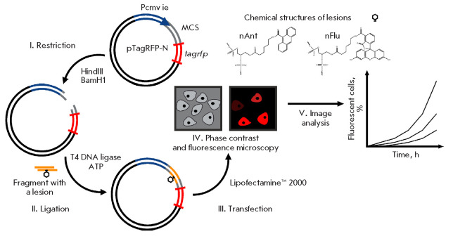
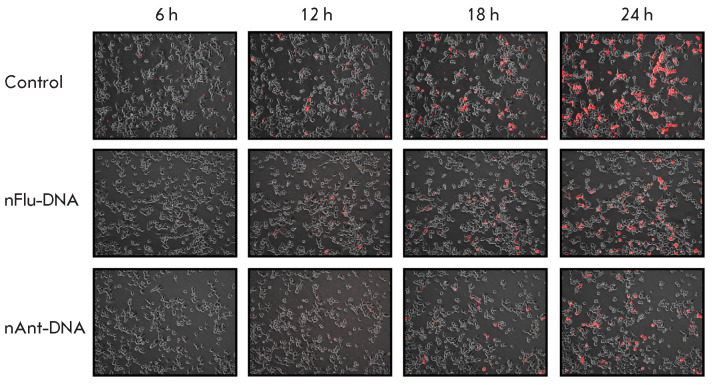
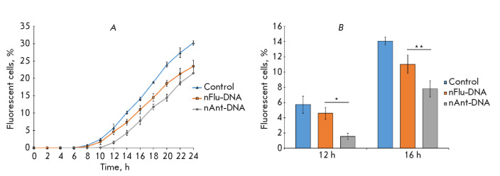
The nucleotide excision repair (NER) is one of the main repair systems present in the cells of living organisms. It is responsible for the removal of a wide range of bulky DNA lesions. We succeeded in developing a method for assessing the efficiency of NER in the cell (ex vivo), which is a method based on the recovery of TagRFP fluorescent protein production through repair of the damage that blocks the expression of the appropriate gene. Our constructed plasmids containing bulky nFlu or nAnt lesions near the tagrfp gene promoter were shown to undergo repair in eukaryotic cells (HEK 293T) and that they can be used to analyze the efficiency of NER ex vivo. A comparative analysis of the time dependence of fluorescent cells accumulation after transfection with nFlu- and nAnt-DNA revealed that there are differences in how efficient their repair by the NER system of HEK 293T cells can be. The method can be used to assess the cell repair status and the repair efficiency of different structural damages.
Keywords: DNA damages; ex vivo methods; nucleotide excision repair.
Copyright ® 2021 National Research University Higher School of Economics.
Figures
Similar articles
-
[DNA Bearing Bulky Fluorescent and Photoreactive Damage in Both Strands as Substrates of the Nucleotide Excision Repair System].Mol Biol (Mosk). 2018 Mar-Apr;52(2):277-288. doi: 10.7868/S0026898418020118. Mol Biol (Mosk). 2018. PMID: 29695696 Russian.
-
DNA with Damage in Both Strands as Affinity Probes and Nucleotide Excision Repair Substrates.Biochemistry (Mosc). 2016 Mar;81(3):263-74. doi: 10.1134/S0006297916030093. Biochemistry (Mosc). 2016. PMID: 27262196
-
Methods for Assessment of Nucleotide Excision Repair Efficiency.Biochemistry (Mosc). 2023 Nov;88(11):1844-1856. doi: 10.1134/S0006297923110147. Biochemistry (Mosc). 2023. PMID: 38105203 Review.
-
Synthetic Lesions with a Fluorescein Carbamoyl Group As Analogs of Bulky Lesions Removable by Nucleotide Excision Repair: A Comparative Study on Properties.Acta Naturae. 2024 Jul-Sep;16(3):74-82. doi: 10.32607/actanaturae.27419. Acta Naturae. 2024. PMID: 39555170 Free PMC article.
-
Novel insights into bulky DNA damage formation and nucleotide excision repair from high-resolution genomics.DNA Repair (Amst). 2023 Oct;130:103549. doi: 10.1016/j.dnarep.2023.103549. Epub 2023 Aug 3. DNA Repair (Amst). 2023. PMID: 37566959 Review.
References
LinkOut - more resources
Full Text Sources
Miscellaneous