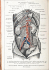
Review: Pelvic nerves - from anatomy and physiology to clinical applications
- PMID: 34707906
- PMCID: PMC8500855
- DOI: 10.1515/tnsci-2020-0184
Review: Pelvic nerves - from anatomy and physiology to clinical applications
Abstract
A prerequisite for nerve-sparing pelvic surgery is a thorough understanding of the topographic anatomy of the fine and intricate pelvic nerve networks, and their connections to the central nervous system. Insights into the functions of pelvic nerves will help to interpret disease symptoms correctly and improve treatment. In this article, we review the anatomy and physiology of autonomic pelvic nerves, including their topography and putative functions. The aim is to achieve a better understanding of the mechanisms of pelvic pain and functional disorders, as well as improve their diagnosis and treatment. The information will also serve as a basis for counseling patients with chronic illnesses. A profound understanding of pelvic neuroanatomy will permit complex surgery in the pelvis without relevant nerve injury.
Keywords: chronic pelvic pain; hypogastric nerves; pelvic neuroanatomy; pelvic neurophysiology; pelvis.
© 2021 Ibrahim Alkatout et al., published by De Gruyter.
Conflict of interest statement
Conflict of interest: Authors state no conflict of interest.
Figures


Similar articles
-
Laparoscopic anatomy of the autonomic nerves of the pelvis and the concept of nerve-sparing surgery by direct visualization of autonomic nerve bundles.Fertil Steril. 2015 Nov;104(5):e11-2. doi: 10.1016/j.fertnstert.2015.07.1138. Epub 2015 Aug 8. Fertil Steril. 2015. PMID: 26260200
-
[Deep infiltrating endometriosis surgical management and pelvic nerves injury].Gynecol Obstet Fertil. 2016 May;44(5):302-8. doi: 10.1016/j.gyobfe.2016.03.007. Epub 2016 Apr 21. Gynecol Obstet Fertil. 2016. PMID: 27118342 Review. French.
-
Anatomy of the female pelvic nerves: a macroscopic study of the hypogastric plexus and their relations and variations.J Anat. 2020 Sep;237(3):487-494. doi: 10.1111/joa.13206. Epub 2020 May 19. J Anat. 2020. PMID: 32427364 Free PMC article.
-
Pelvic Neuroanatomy: An Overview of Commonly Encountered Pelvic Nerves in Gynecologic Surgery.J Minim Invasive Gynecol. 2021 Feb;28(2):178. doi: 10.1016/j.jmig.2020.06.005. Epub 2020 Jun 12. J Minim Invasive Gynecol. 2021. PMID: 32540500 Review.
-
Laparoscopic pelvic autonomic nerve-preserving surgery for patients with lower rectal cancer after chemoradiation therapy.Ann Surg Oncol. 2007 Apr;14(4):1285-7. doi: 10.1245/s10434-006-9052-6. Ann Surg Oncol. 2007. PMID: 17235719 Clinical Trial.
Cited by
-
Automatic muscle impedance and nerve analyzer (AMINA) as a novel approach for classifying bioimpedance signals in intraoperative pelvic neuromonitoring.Sci Rep. 2024 Jan 5;14(1):654. doi: 10.1038/s41598-023-50504-7. Sci Rep. 2024. PMID: 38182695 Free PMC article.
-
The distribution of the inferior hypogastric plexus in female pelvis.J Med Life. 2022 Jun;15(6):784-791. doi: 10.25122/jml-2022-0145. J Med Life. 2022. PMID: 35928357 Free PMC article.
-
Emergency treatment of pelvic ring injuries: state of the art.Arch Orthop Trauma Surg. 2024 Oct;144(10):4525-4539. doi: 10.1007/s00402-024-05447-7. Epub 2024 Jul 6. Arch Orthop Trauma Surg. 2024. PMID: 38970673 Free PMC article. Review.
-
Peripheral Nerve Blocks for Cesarean Delivery Analgesia: A Narrative Review.Medicina (Kaunas). 2023 Nov 4;59(11):1951. doi: 10.3390/medicina59111951. Medicina (Kaunas). 2023. PMID: 38004000 Free PMC article. Review.
-
Intraoperative pelvic neuromonitoring based on bioimpedance signals: a new method analyzed on 30 patients.Langenbecks Arch Surg. 2024 Aug 3;409(1):237. doi: 10.1007/s00423-024-03403-y. Langenbecks Arch Surg. 2024. PMID: 39096391 Free PMC article.
References
-
- Langley JN. On the union of cranial autonomic (visceral) fibres with the nerve cells of the superior cervical ganglion. J Physiol. 1898;23(3):240. - PMC - PubMed
- Langley JN. On the union of cranial autonomic (visceral) fibres with the nerve cells of the superior cervical ganglion. J Physiol. 1898;23(3):240. - PMC - PubMed
-
- Langley JN. The autonomic nervous system. vol. 1, 4th ed. Cambridge, MA: Heffer and sons; 1921.
- Langley JN. The autonomic nervous system. vol. 1, 4th ed. Cambridge, MA: Heffer and sons. 1921.
-
- Okabayashi H. Radical abdominal hysterectomy for cancer of the cervix uteri. Modification of the Takayama operation. Surg Gynecol Obstet. 1921;33:335–43.
- Okabayashi H. Radical abdominal hysterectomy for cancer of the cervix uteri. Modification of the Takayama operation. Surg Gynecol Obstet. 1921;33:335–43.
-
- Muallem MZ, Diab Y, Sehouli J, Fujii S. Nerve-sparing radical hysterectomy: steps to standardize surgical technique. Int J Gynecol Cancer. 2019;29:1203–8. - PubMed
- Muallem MZ, Diab Y, Sehouli J, Fujii S. Nerve-sparing radical hysterectomy: steps to standardize surgical technique. Int J Gynecol Cancer. 2019;29:1203–8. - PubMed
-
- Sakamoto S, Takizawa K. 20 An improved radical hysterectomy with fewer urological complications and with no loss of therapeutic results for invasive cervical cancer. Baillière’s Clin Obstet Gynaecol. 1988;2(4):953–62. - PubMed
- Sakamoto S, Takizawa K. 20 An improved radical hysterectomy with fewer urological complications and with no loss of therapeutic results for invasive cervical cancer. Baillière’s Clin Obstet Gynaecol. 1988;2(4):953–62. - PubMed
Publication types
LinkOut - more resources
Full Text Sources
