. 2021 Oct 25;7(4):00397-2021.
doi: 10.1183/23120541.00397-2021. eCollection 2021 Oct.

The utility of gallium-68 DOTATOC PET/CT in lymphangioleiomyomatosis

Affiliations

The utility of gallium-68 DOTATOC PET/CT in lymphangioleiomyomatosis

Brian Gaffney et al. ERJ Open Res. .

Abstract

Somatostatin receptor functional imaging is of limited utility as an imaging biomarker in LAM, but other PET/CT modalities may be of use https://bit.ly/3l6BVZp.


Figures

FIGURE 1
FIGURE 1
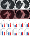
Qualitative and quantitative assessment of 68Ga-DOTA-TyI3-octreotide (DOTATOC) positron emission tomography (PET)/computed tomography (CT) in lymphangioleiomyomatosis (LAM). a) Axial thoracic CT image at the level of the carina in a patient with mild LAM (forced expiratory volume in 1 s (FEV1): 99% of predicted; diffusing capacity of the lung for carbon monoxide (DLCO): 88% of predicted) demonstrates occasional scattered thin-walled cysts. c) No increased tracer uptake evident on the axial fused 68Ga-DOTATOC PET/CT image (maximum standardised uptake value (SUVmax) window range 0–5). b) A separate patient with severe LAM (FEV1: 36% of predicted; DLCO: 25% of predicted) has innumerable cysts demonstrated on this axial thoracic CT image at the level of the aortic arch. d) No increased pulmonary tracer uptake evident on 68Ga-DOTATOC PET/CT. Dashed line: sample region of interest demonstrated on PET images panels c and d. e) Semiquantitative SUVmax measures of pulmonary 68Ga-DOTATOC uptake in lungs of patients with LAM and controls. f) Target to background ratio for each lobe calculated by dividing the lobar SUVmax by the mediastinal blood pool SUVmax in lungs of patients with LAM and controls. Each dot represents an individual patient. Data are presented as mean±sd. LAM, n=4; control; n=4. ns: nonsignificant. *: p<0.05, unpaired Student's t-test.

References

    1. Henske EP, McCormack FX. Lymphangioleiomyomatosis – a wolf in sheep's clothing. J Clin Invest 2012: 122: 3807–3816. doi:10.1172/JCI58709 - DOI - PMC - PubMed
    1. Young LR, Lee HS, Inoue Y, et al. . Serum VEGF-D concentration as a biomarker of lymphangioleiomyomatosis severity and treatment response: a prospective analysis of the Multicenter International Lymphangioleiomyomatosis Efficacy of Sirolimus (MILES) trial. Lancet Respir Med 2013: 1: 445–452. doi:10.1016/S2213-2600(13)70090-0 - DOI - PMC - PubMed
    1. Young LR, Franz DN, Nagarkatte P, et al. . Utility of [18F]2-fluoro-2-deoxyglucose-PET in sporadic and tuberous sclerosis-associated lymphangioleiomyomatosis. Chest 2009: 136: 926–933. doi:10.1378/chest.09-0336 - DOI - PMC - PubMed
    1. Johnson SR, Tattersfield AE. Clinical experience of lymphangioleiomyomatosis in the UK. Thorax 2000: 55: 1052–1057. doi:10.1136/thorax.55.12.1052 - DOI - PMC - PubMed
    1. Ryu JH, Doerr CH, Fisher SD, et al. . Chylothorax in lymphangioleiomyomatosis. Chest 2003: 123: 623–627. doi:10.1378/chest.123.2.623 - DOI - PubMed

LinkOut - more resources