Review
. 2022 Jan 28;42(1):BSR20211754.
doi: 10.1042/BSR20211754.

Epithelial-mesenchymal transition and its transcription factors

Affiliations
Review

Epithelial-mesenchymal transition and its transcription factors

Pallabi Debnath et al. Biosci Rep. .

Abstract

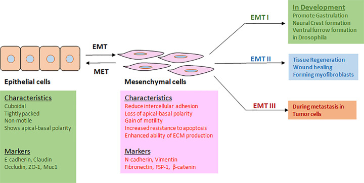
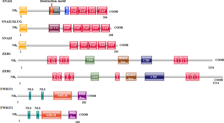
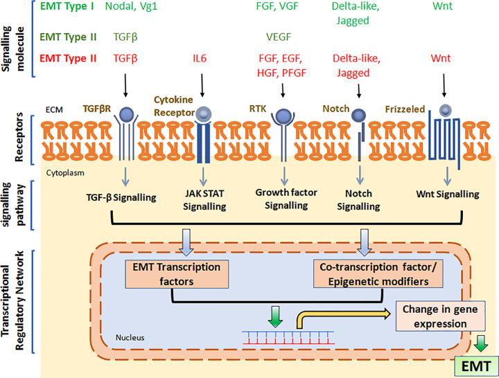
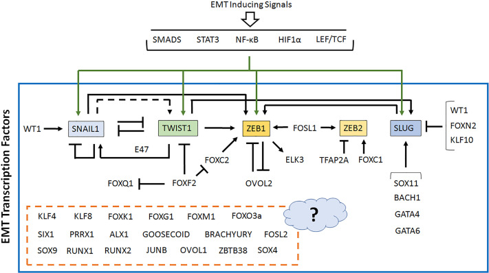
Epithelial-mesenchymal transition or EMT is an extremely dynamic process involved in conversion of epithelial cells into mesenchymal cells, stimulated by an ensemble of signaling pathways, leading to change in cellular morphology, suppression of epithelial characters and acquisition of properties such as enhanced cell motility and invasiveness, reduced cell death by apoptosis, resistance to chemotherapeutic drugs etc. Significantly, EMT has been found to play a crucial role during embryonic development, tissue fibrosis and would healing, as well as during cancer metastasis. Over the years, work from various laboratories have identified a rather large number of transcription factors (TFs) including the master regulators of EMT, with the ability to regulate the EMT process directly. In this review, we put together these EMT TFs and discussed their role in the process. We have also tried to focus on their mechanism of action, their interdependency, and the large regulatory network they form. Subsequently, it has become clear that the composition and structure of the transcriptional regulatory network behind EMT probably varies based upon various physiological and pathological contexts, or even in a cell/tissue type-dependent manner.

Keywords: Epithelial mesenchymal transition; Transcription factor; gene expression regulation; transcriptional regulatory network.


Conflict of interest statement

The authors declare that there are no competing interests associated with the manuscript.

Figures

Figure 1
Figure 1. EMT: characteristics, markers, and contexts
EMT is activated in different physiological and pathological contexts, thereby facilitating cellular movement. It is associated with both morphological and characteristics’ changes.
Figure 2
Figure 2. Structure of master regulators of EMT
Schematic depiction of EMT master regulators and their respective domains with their comparative size; ZEB2 being the largest and TWIST2 being smallest. SNAI1 and SNAI2 have zinc fingers in their C-terminus, whereas ZEB1 and ZEB2 have zinc fingers on both sides.
Figure 3
Figure 3. EMT overview
EMT is induced by a variety of signaling molecules, which stimulate cognate receptors on the cell surface and thereby activate downstream signaling cascade, leading to activation of EMT TFs and associated co-regulators and epigenetic regulators. This subsequently turns ‘on’ or ‘off’ specific genes. The altered transcriptome and proteome further supports the transition.
Figure 4
Figure 4. Transcriptional regulatory network in EMT
EMT is regulated by many TFs, expression of which are dependent on each other to a large extent, thereby creating a complex transcriptional regulatory network. The master regulators of EMT are shown within colored boxes, while all other EMT TFs are shown as it is. For some EMT TFs, no information regarding their cross-regulation is known, so these are kept within a broken box attached with a question mark. The transcriptional regulatory network is derived from currently available literature (please see text for details) and include all different cell/tissue types as well as different EMT contexts.
Figure 5
Figure 5. Genetic alterations of EMT TF genes
TCGA data were analyzed for the presence of (A) SSMs and (B) CNV within the set of EMT TFs discussed in this review. Our analysis revealed ZEB2, SOX11, and ZEB1 to be the most frequently mutated EMT TFs (SSMs), whereas TCF3, TWIST2, KLF10, and SOX9 to be the EMT TFs with most frequently altered CNV cases.
Figure 6
Figure 6. The EMT inhibitors and their targets
Small molecule inhibitors are being used to suppress the EMT process. They target various key components of EMT, including the EMT TFs.

References

    1. Lee J.M., Dedhar S., Kalluri R. and Thompson E.W. (2006) The epithelial-mesenchymal transition: new insights in signaling, development, and disease. J. Cell Biol. 172, 973–981 10.1083/jcb.200601018 - DOI - PMC - PubMed
    1. Yang J. and Weinberg R.A. (2008) Epithelial-mesenchymal transition: at the crossroads of development and tumor metastasis. Dev. Cell 14, 818–829, https://www.sciencedirect.com/science/article/pii/S1534580708002098 10.1016/j.devcel.2008.05.009 - DOI - PubMed
    1. Kalluri R. and Weinberg R.A. (2009) The basics of epithelial-mesenchymal transition. J. Clin. Invest. 119, 1420–1428 10.1172/JCI39104 - DOI - PMC - PubMed
    1. Thiery J.P., Acloque H., Huang R.Y.J. and Nieto M.A. (2009) Epithelial-mesenchymal transitions in development and disease. Cell 139, 871–890 https://www.sciencedirect.com/science/article/pii/S0092867409014196 10.1016/j.cell.2009.11.007 - DOI - PubMed
    1. Lamouille S., Xu J. and Derynck R. (2014) Molecular mechanisms of epithelial-mesenchymal transition. Nat. Rev. Mol. Cell Biol. 15, 178–196 10.1038/nrm3758 - DOI - PMC - PubMed

Publication types

MeSH terms