

Structural insights into glucocorticoid receptor function
- PMID: 34709368
- PMCID: PMC9274455
- DOI: 10.1042/BST20210419
Structural insights into glucocorticoid receptor function
Abstract
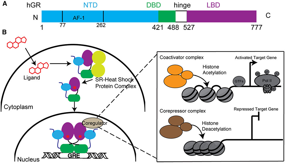
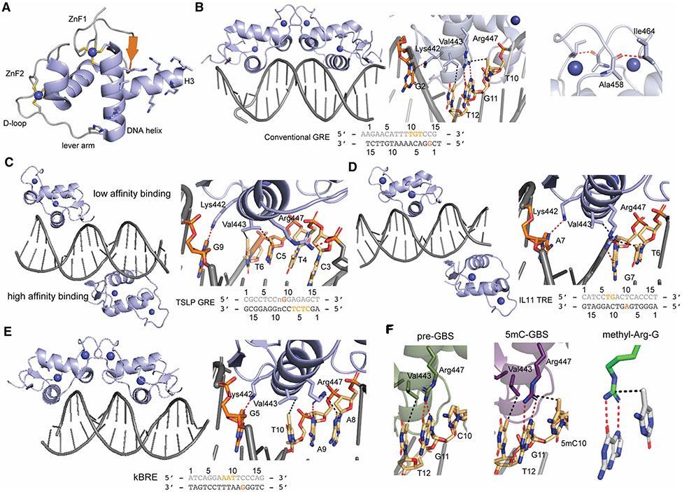
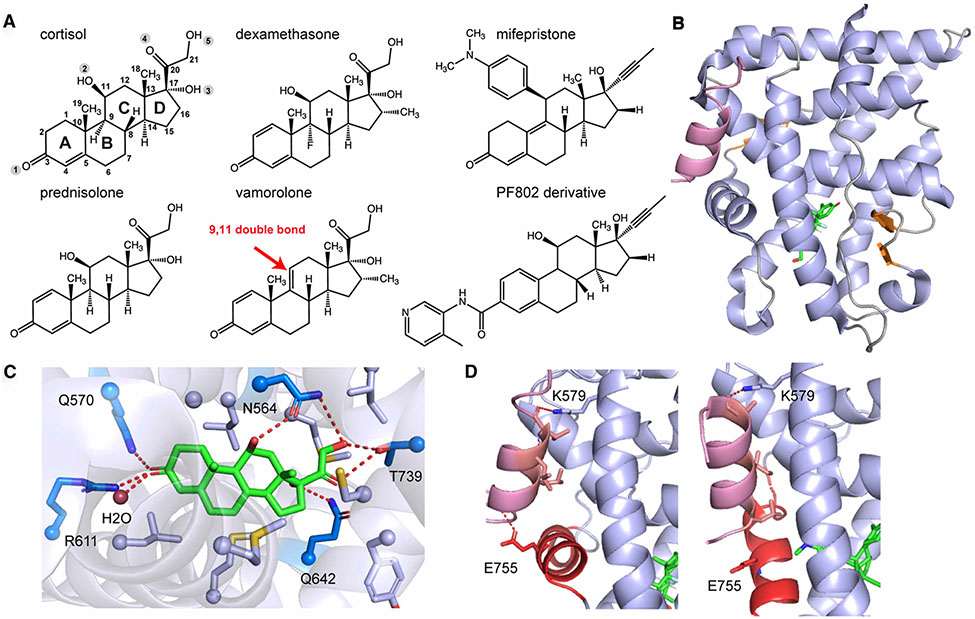
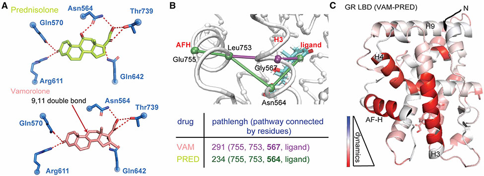
The glucocorticoid receptor (GR) is a steroid hormone-activated transcription factor that binds to various glucocorticoid response elements to up- or down- regulate the transcription of thousands of genes involved in metabolism, development, stress and inflammatory responses. GR consists of two domains enabling interaction with glucocorticoids, DNA response elements and coregulators, as well as a large intrinsically disordered region that mediates condensate formation. A growing body of structural studies during the past decade have shed new light on GR interactions, providing a new understanding of the mechanisms driving context-specific GR activity. Here, we summarize the established and emerging mechanisms of action of GR, primarily from a structural perspective. This minireview also discusses how the current state of knowledge of GR function may guide future glucocorticoid design with an improved therapeutic index for different inflammatory disorders.
Keywords: DNA binding domain; glucocorticoid receptor; ligand binding domain; transactivation; transrepression.
© 2021 The Author(s). Published by Portland Press Limited on behalf of the Biochemical Society.
Conflict of interest statement
Competing Interests
The authors declare that there are no competing interests associated with the manuscript.
Figures
Similar articles
-
Glucocorticoid receptor condensates link DNA-dependent receptor dimerization and transcriptional transactivation.Proc Natl Acad Sci U S A. 2021 Jul 27;118(30):e2024685118. doi: 10.1073/pnas.2024685118. Proc Natl Acad Sci U S A. 2021. PMID: 34285072 Free PMC article.
-
First High-Resolution Crystal Structures of the Glucocorticoid Receptor Ligand-Binding Domain-Peroxisome Proliferator-Activated γ Coactivator 1-α Complex with Endogenous and Synthetic Glucocorticoids.Mol Pharmacol. 2019 Oct;96(4):408-417. doi: 10.1124/mol.119.116806. Epub 2019 Aug 7. Mol Pharmacol. 2019. PMID: 31391291 Free PMC article.
-
The structural basis of direct glucocorticoid-mediated transrepression.Nat Struct Mol Biol. 2013 Jan;20(1):53-8. doi: 10.1038/nsmb.2456. Epub 2012 Dec 9. Nat Struct Mol Biol. 2013. PMID: 23222642 Free PMC article.
-
How glucocorticoid receptors modulate the activity of other transcription factors: a scope beyond tethering.Mol Cell Endocrinol. 2013 Nov 5;380(1-2):41-54. doi: 10.1016/j.mce.2012.12.014. Epub 2012 Dec 23. Mol Cell Endocrinol. 2013. PMID: 23267834 Review.
-
New insights into the anti-inflammatory mechanisms of glucocorticoids: an emerging role for glucocorticoid-receptor-mediated transactivation.Endocrinology. 2013 Mar;154(3):993-1007. doi: 10.1210/en.2012-2045. Epub 2013 Feb 5. Endocrinology. 2013. PMID: 23384835 Review.
Cited by
-
Glucocorticoid Treatment in Acute Respiratory Distress Syndrome: An Overview on Mechanistic Insights and Clinical Benefit.Int J Mol Sci. 2023 Jul 28;24(15):12138. doi: 10.3390/ijms241512138. Int J Mol Sci. 2023. PMID: 37569514 Free PMC article. Review.
-
α1-Antitrypsin Binds to the Glucocorticoid Receptor with Anti-Inflammatory and Antimycobacterial Significance in Macrophages.J Immunol. 2022 Nov 1;209(9):1746-1759. doi: 10.4049/jimmunol.2200227. Epub 2022 Sep 26. J Immunol. 2022. PMID: 36162872 Free PMC article.
-
Sex hormones and allergies: exploring the gender differences in immune responses.Front Allergy. 2025 Jan 7;5:1483919. doi: 10.3389/falgy.2024.1483919. eCollection 2024. Front Allergy. 2025. PMID: 39840271 Free PMC article. Review.
-
Transcriptomic and Chromatin Landscape Analysis Reveals That Involvement of Pituitary Level Transcription Factors Modulate Incubation Behaviors of Magang Geese.Genes (Basel). 2023 Mar 28;14(4):815. doi: 10.3390/genes14040815. Genes (Basel). 2023. PMID: 37107573 Free PMC article.
-
BGATT-GR: accurate identification of glucocorticoid receptor antagonists based on data augmentation combined with BiGRU-attention.Sci Rep. 2025 Jul 1;15(1):21402. doi: 10.1038/s41598-025-05839-8. Sci Rep. 2025. PMID: 40595974 Free PMC article.
References
Publication types
MeSH terms
Substances
Grants and funding
LinkOut - more resources
Full Text Sources
Miscellaneous