Review
. 2021 Oct 29;478(20):3791-3805.
doi: 10.1042/BCJ20210412.

Meiosis initiation: a story of two sexes in all creatures great and small

Affiliations
Review

Meiosis initiation: a story of two sexes in all creatures great and small

Ieng Fong Sou et al. Biochem J. .

Abstract

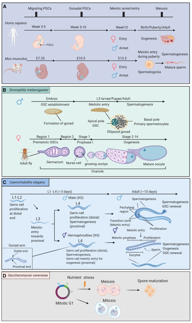
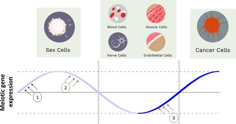
Meiosis facilitates diversity across individuals and serves as a major driver of evolution. However, understanding how meiosis begins is complicated by fundamental differences that exist between sexes and species. Fundamental meiotic research is further hampered by a current lack of human meiotic cells lines. Consequently, much of what we know relies on data from model organisms. However, contextualising findings from yeast, worms, flies and mice can be challenging, due to marked differences in both nomenclature and the relative timing of meiosis. In this review, we set out to combine current knowledge of signalling and transcriptional pathways that control meiosis initiation across the sexes in a variety of organisms. Furthermore, we highlight the emerging links between meiosis initiation and oncogenesis, which might explain the frequent re-expression of normally silent meiotic genes in a variety of human cancers.

Keywords: TEX12; cancer; cancer testis antigen; meiosis; meiosis initiation; model organisms.


Conflict of interest statement

The authors declare that there are no competing interests associated with the manuscript.

Figures

Figure 1.
Figure 1.. The timing of meiotic entry in distinct species and sexes.
(A) In Homo sapiens and Mus musculus. Meiotic entry is largely conserved. In general, PGCs migrate through the midgut and arrive in the fetal gonads, where meiosis occurs in females and mitosis arrests in males. (B) In Drosophila melanogaster germ cells are derived from GSCs. Meiosis occurs in L3 larval instar stage in the male, GSCs shift from the apical pole to differentiate into primary spermatocytes in the basal pole, whereas GSCs enter prophase I within region 2 of germarium in adult female ovariole. (C) In Caenorhabditis elegans the dioecious hermaphroditic and male worms both enter meiosis during the L3 stage, GSCs shift from the distal to the proximal end as meiosis progresses. During L4 stage, male worms undergo spermatogenesis similarly to hermaphroditic worms but in hermaphrodites a subgroup of germ cells enter meiosis for oogenesis. With the renewal of GSCs, oogenesis and spermatogenesis can be maintained throughout adulthood. (D) Saccharomyces cerevisiae reproduces asexually upon nutrient starvation mitosis arrests at G1 and meiotic entry is triggered.
Figure 2.
Figure 2.. Expression of genes understood to be exclusively meiotic is assumed tightly regulated and silenced in healthy somatic cells.
However, we still lack detailed mechanistic understanding of the machinery that turns on meiotic gene expression during meiosis initiation (1), turns expression off during the meiotic-to-mitotic transition (2), and (3) the factors responsible for aberrant reactivation of meiotic gene expression in, for example, cancer cells.
Figure 3.
Figure 3.. Unanswered questions about meiotic gene activation.
(1) At the DNA level, interactions between the gene promoter (pink) and enhancer (purple) regions controls recruitment of transcription regulating machinery including transcription factors/repressors and their cofactors. What are the cell type specific meiotic promoter binding proteins and how do they regulate exclusively meiotic expression? (2) The availability of each meiotic promoter binding protein (shown as (1)) is affected by a network of specific cofactors (rectangle), kinases (star), phosphatases (diamond), E3s (triangle), deubiquitinating enzymes (oval) and others (trapezoid). What is the complete network of these meiotic expression master regulators? (3) Gene expression is affected by distal enhancers (purple) and the 3D genome architecture which controls their ability to interact with gene promoters (pink) to facilitate the recruitment of transcriptional machinery. What are the cell type specific distal enhancers of meiotic genes? (4) At a higher level of genome compaction large topologically associated domains (TADs) become the units of chromatin and a position of the gene within the TADs can affect its expression. What are the cell type specific TAD domain positions of meiotic genes and what role does the TAD positioning play in regulating the expression of meiotic genes?

References

    1. Mitchell, A.P. (1994) Control of meiotic gene expression in Saccharomyces cerevisiae. Microbiol. Rev. 58, 56–70 10.1128/mr.58.1.56-70.1994 - DOI - PMC - PubMed
    1. Covitz, P.A. and Mitchell, A.P. (1993) Repression by the yeast meiotic inhibitor RME1. Genes Dev. 7, 1598–1608 10.1101/gad.7.8.1598 - DOI - PubMed
    1. Chu, S., DeRisi, J., Eisen, M., Mulholland, J., Botstein, D., Brown, P.O.et al. (1998) The transcriptional program of sporulation in budding yeast. Science 282, 699–705 10.1126/science.282.5389.699 - DOI - PubMed
    1. Honigberg, S.M. and Purnapatre, K. (2003) Signal pathway integration in the switch from the mitotic cell cycle to meiosis in yeast. J. Cell Sci. 116, 2137–2147 10.1242/jcs.00460 - DOI - PubMed
    1. Nachman, I., Regev, A. and Ramanathan, S. (2007) Dissecting timing variability in yeast meiosis. Cell 131, 544–556 10.1016/j.cell.2007.09.044 - DOI - PubMed

Publication types

MeSH terms