. 2021 Jan-Dec:20:15330338211045831.
doi: 10.1177/15330338211045831.

CBX2 Induces Glioma Cell Proliferation and Invasion Through the Akt/PI3K Pathway

Affiliations

CBX2 Induces Glioma Cell Proliferation and Invasion Through the Akt/PI3K Pathway

Le Wang et al. Technol Cancer Res Treat. 2021 Jan-Dec.

Abstract

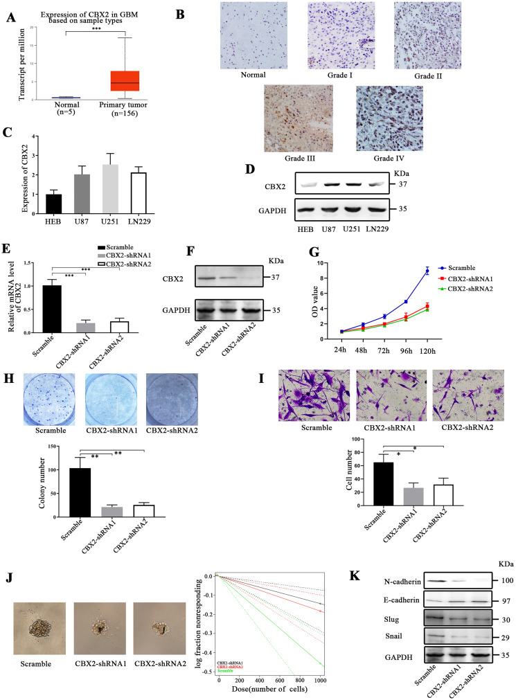
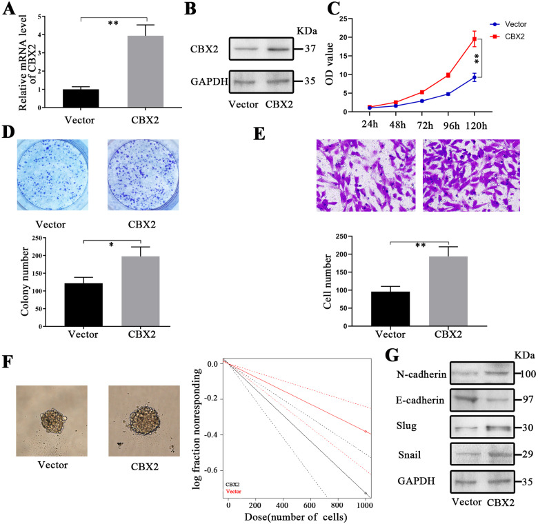
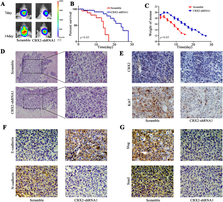
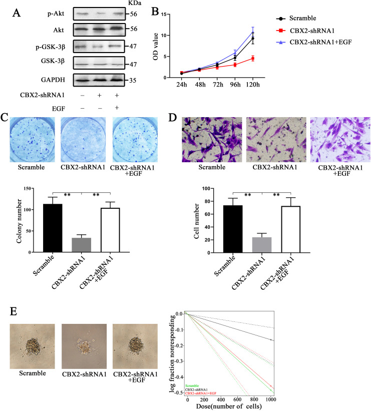
Glioma is the most common primary intracranial tumor. Abnormal expression of CBX2 (ChromoBox2) is associated with tumorigenesis and tumor development. TCGA data in UALCAN showed that CBX2 was overexpressed in glioma tissue. To confirm the role of CBX2 in glioma, we regulated the level of CBX2 and conducted colony formation, Transwell, and CCK-8 assays to verify the effect of CBX2. The results showed that CBX2 knockdown reduced glioma cell proliferation and invasion and that the cells were less tumorigenic. CBX2 overexpression induced glioma cell proliferation and invasion and glioma stem cell self-renewal. The animal experiments showed that CBX2 knockdown inhibited glioma growth and improved survival time. CBX2 knockdown inhibited activation of the Akt/PI3K pathway. epidermal growth factor rescued the effects of CBX2. CBX2 could induce the growth and invasion of glioma cells via the Akt/PI3K pathway.

Keywords: Akt; CBX2; PI3K; glioma.


Conflict of interest statement

Declaration of Conflicting Interests: The authors declared no potential conflicts of interest with respect to the research, authorship, and/or publication of this article.

Figures

Figure 1.
Figure 1.
Downregulation of CBX2 inhibited glioma cell proliferation, invasion, and self-renewal. (A) The data (UALCAN, http://ualcan.path.uab.edu/index.html) showed that the expression of CBX2 was higher than that in normal brain tissue (P < .001). (B) The level of CBX2 was higher in glioma tissues. And with the increase of glioma grade, the expression of CBX2 increased, the expression of CBX2 in Grade IV glioma tissues was the highest. (C and D) The expression of CBX2 was analyzed in glioma cell lines by RT-PCR and western blotting. (E and F) CBX2-shRNA transfection efficiencies were verified by RT-PCR and western blotting (P < .001). (G and H) CCK-8 and colony formation assays demonstrated that CBX2-shRNA-mediated knockdown of CBX2 decreased the proliferation of glioma cells (P < .01). (I) Transwell assays showed that CBX2 downregulation reduced invasion compared to that in the scramble group. (J) Sphere formation analysis showed that the downregulation of CBX2 decreased the glioma stem cell frequencies (P < .05). (K) Knockdown of CBX2 reduced the levels of N-cadherin, slug, and snail and increased the level of E-cadherin.
Figure 2.
Figure 2.
Upregulation of CBX2 induced glioma cell proliferation, invasion, and self-renewal. (A and B) CBX2 plasmid transfection efficiencies were verified by RT-PCR and western blotting (P < .01). (C and D) CCK-8 and colony formation assays demonstrated that CBX2 plasmid-mediated overexpression of CBX2 induced the proliferation of glioma cells (P < .01, P < .05). (E) Transwell assays showed that CBX2 upregulation induced invasion compared to that of the scramble group (P < .01). (F) The glioma stem cell frequency was increased following transfection with the CBX2 plasmid, and the effects were analyzed by sphere formation assays. (G) The upregulation of CBX2 reduced the level of E-cadherin and increased the expression of N-cadherin, slug, and snail.
Figure 3.
Figure 3.
Knockdown of CBX2 repressed tumor growth in vivo. (A) Fluc bioluminescence images showing the tumors in the nude mouse xenograft model. (B) Overall survival analysis demonstrated that CBX2 downregulation improved the survival time (P < .05). (C) The mouse body weights were evaluated. (D) Images of HE-stained tumor tissue. (E) The expression of CBX2 and Ki67 was analyzed by IHC staining. (F and G) The expression of EMT markers, E-cadherin, N-cadherin, slug, and slug were detected with IHC.
Figure 4.
Figure 4.
Knockdown of CBX2 inhibited glioma cell proliferation and invasion and glioma stem cell self-renewal via the downregulation of the Akt/PI3K pathway. (A) Knockdown of CBX2 inhibited the levels of p-Akt and p-GSK-3β, reducing the activity of the Akt/PI3K pathway. The use of EGF, which is an Akt/PI3K pathway agonist, increased the levels of p-Akt and p-GSK-3β. (B and C) CCK-8 and colony formation assays showed that EGF rescued the proliferation of glioma cells. (D) Sphere formation assays showed that the cells in the CBX2-shRNA1 + EGF group were more tumorigenic than those in the CBX2-shRNA1 group (P < .05).

References

    1. McNeill KA. Epidemiology of brain tumors. Neurol Clin. 2016;34(4):981‐998. - PubMed
    1. Wen PY, Reardon DA. Neuro-oncology in 2015: progress in glioma diagnosis, classification and treatment. Nat Rev Neurol. 2016;12(2):69‐70. - PubMed
    1. Soomro SH, Ting LR, Qing YY, Ren M. Molecular biology of glioblastoma: classification and mutational locations. J Pak Med Assoc. 2017;67(9):1410‐1414. - PubMed
    1. Gil J, Bernard D, Martínez D, Beach D. Polycomb CBX7 has a unifying role in cellular lifespan. Nat Cell Biol. 2004;6(1):67‐72. - PubMed
    1. Sun D, Cao X, Wang C. Polycomb chromobox Cbx2 enhances antiviral innate immunity by promoting Jmjd3-mediated demethylation of H3K27 at the Ifnb promoter. Protein Cell. 2019;10(4):285‐294. - PMC - PubMed

Publication types

MeSH terms