. 2021 Oct 28;7(1):318.
doi: 10.1038/s41420-021-00720-9.

miR548ai antagonism attenuates exosome-induced endothelial cell dysfunction

Affiliations

miR548ai antagonism attenuates exosome-induced endothelial cell dysfunction

Xiujie Xie et al. Cell Death Discov. .

Erratum in

Abstract

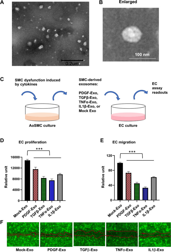
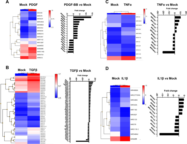
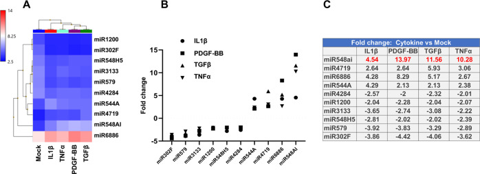
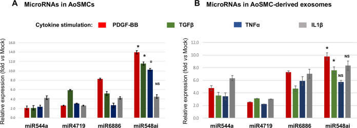
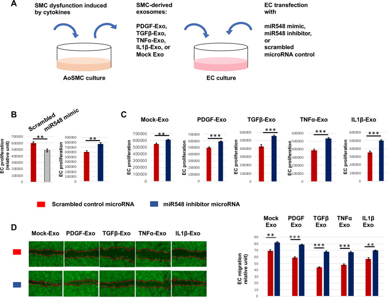
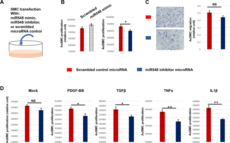
Endothelial cell (EC) and smooth muscle cell (SMC) are major cell types adjacent in the vascular wall. Recent progress indicates that their communication is crucial for vascular homeostasis and pathogenesis. In particular, dysfunctional (proliferative) SMCs through exosomes can induce EC dysfunction (impaired growth). The current study suggests that miR548ai, a rarely known microRNA, may provide a molecular target for protection against SMC/exosome-induced EC dysfunction. We performed microarray profiling of microRNAs of dysfunctional human primary aortic SMCs induced by different cytokines (PDGF-BB, TGFβ1, TNFα, IL1β). Among the microRNAs commonly upregulated by these cytokines, miR548ai showed the most robust changes, as also validated through quantitative PCR. This cytokine-induced miR548ai upregulation was recapitulated in the qPCR determination of SMC-derived exosomal microRNAs. Consistent with SMC-to-EC communication, the exosomes extracted from cytokine-stimulated SMCs impaired human EC proliferation and migration. Of particular interest, this SMC exosomal impingement on ECs was countered by transfection of miR548ai inhibitor microRNA into ECs. Furthermore, the miR548ai inhibitor transfected into SMCs attenuated SMC dysfunction/proliferation. Thus, these results identify miR548ai as a novel target; namely, miR548ai inhibitor mitigates EC dysfunction induced by exosomes derived from dysfunctional SMCs. This new knowledge may aid the future development of microRNA-based treatment of vascular disorders.


Conflict of interest statement

The authors declare no competing interests.

Figures

Fig. 1
Fig. 1. Inhibitory effect of dysfunctional SMC-derived exosomes on EC proliferation and migration.
Human primary aortic smooth muscle cells (AoSMCs) were starved in basal medium (no FBS) and then treated for 24 h without (Mock control) or with a cytokine (50 ng/ml PDGF-BB, 20 ng/ml TGFβ1, 20 ng/ml TNFα, or 10 ng/ml IL1β). Exosomes extracted from a conditioned medium were added to human umbilical vascular ECs cultured in full medium and incubated for 48 or 24 h prior to measurements of proliferation and migration, respectively. Data are presented as mean ± SD (n = 3 replicates). ***P < 0.001 (compared to any of the cytokine conditions), as analyzed with one‐way ANOVA followed by Bonferroni post hoc test. A TEM image of SMC-derived exosomes. Purified exosomes were resuspended in PBS. Scale bar: 200 nm. B Enlarged image of a single exosome. Scale bar: 100 nm. C Schematic of the experimental design for this figure. D EC proliferation (Cell Titer-Glo assay). E EC migration (scratch assay). F Representative images showing cell-free gaps 24 h after scratch. Migration was measured as an increase in cell-reoccupied gap space 24 h (vs. 0 h) after scratching. Calcein was used to render cells fluorescent for imaging at the end of the 24 h experiment.
Fig. 2
Fig. 2. Profiles of SMC-derived, up- or down-regulated microRNAs after cytokine stimulation.
Starved AoSMCs were cultured for 48 h without (Mock control) or with a cytokine (A: PDGF-BB, B: TGFβ1, C: TNFα, D: IL1β). Total RNA extracted from AoSMCs was used for microarray and microRNA profiling. Shown are heatmaps and fold-change profiles of microRNAs that meet the criterion of log2-fold change >2 (cytokine vs. Mock).
Fig. 3
Fig. 3. microRNAs that are commonly up- or down-regulated by all four cytokines.
The AoSMC-derived microRNAs that were commonly up- or down-regulated by all four cytokines were selected (from the data in Fig. 2), and the data are presented as heatmap (A), plot (B), or table of fold changes (C).
Fig. 4
Fig. 4. qRT-PCR determination of cytokine-stimulated upregulation of SMC-derived exosomal microRNAs.
AoSMC culture, cytokine stimulation, and exosome isolation were performed as described in Fig. 1. qRT-PCR was performed to detect cytokine-induced level changes of the four microRNAs selected in Fig. 3. Levels of microRNAs in either cytokine-stimulated AoSMCs or AoSMC-derived exosomes were measured, as shown in A and B, respectively. The relative expression refers to microRNA fold change with (vs. without) cytokine stimulation. Data are presented as mean ± SD (n = 3 replicates). *P < 0.05, miR548ai compared to any of the other three microRNAs (between two bars of the same color), as analyzed with one‐way ANOVA followed by Bonferroni post hoc test; NS not significant.
Fig. 5
Fig. 5. Effect of miR548ai inhibitor on EC proliferation and migration.
A Schematic of the experimental design for this figure. B EC proliferation in full medium. ECs cultured in the full medium were transfected with a miR548ai mimic or inhibitor or scrambled microRNA (control) prior to Cell Titer-Glo assay. C and D EC proliferation and migration (respectively) in the presence of SMC-derived exosomes. Exosomes were isolated from the conditioned media of AoSMCs that were starved and then stimulated with a cytokine (same concentration as indicated in Fig. 1). The exosomes and miR548ai inhibitor (or scrambled microRNA) were added to ECs. Migration was measured as an increase in cell-reoccupied gap space 24 h (vs. 0 h) after scratching. Calcein was used to render cells fluorescent for imaging at the end of the 24 h experiment. Data are presented as mean ± SD (n = 3 replicates). Student t-test: **P < 0.01, ***P < 0.001.
Fig. 6
Fig. 6. Effect of miR548ai inhibitor on AoSMC proliferation and migration.
A Schematic of the experimental design for this figure. B SMC proliferation in full medium. AoSMCs cultured in the full medium were transfected with miR548ai mimic or inhibitor or scrambled microRNA (control) prior to CellTiter-Glo assay. C SMC migration in full medium. AoSMCs cultured in the full medium were transfected with miR548ai inhibitor or scrambled microRNA (control) prior to the Transwell migration assay. D SMC proliferation in defined medium. Starved AoSMCs were transfected with miR548ai inhibitor or scrambled control prior to stimulation with PDGF-BB, TGFβ1, TNFα, or IL1β (same concentrations as indicated in Fig. 1). Data are presented as mean ± SD (n = 3 replicates). Student t-test: *P < 0.05, **P < 0.01; NS not significant.
Fig. 7
Fig. 7. Schematic working model for the effect of miR548ai antagonism on EC and SMC dysfunction.
Whereas exosomes (Exo) derived from dysfunctional SMCs induce EC dysfunction, miR548ai inhibitor mitigates this effect. miR548ai mimic exacerbates, and miR548ai inhibitor attenuates EC and SMC dysfunction.

References

    1. Lilly B. We have contact: endothelial cell–smooth muscle cell interactions. Physiology. 2014;29:234–41. doi: 10.1152/physiol.00047.2013. - DOI - PubMed
    1. Lutter S, Xie S, Tatin F, Makinen T. Smooth muscle–endothelial cell communication activates Reelin signaling and regulates lymphatic vessel formation. J Cell Biol. 2012;197:837–49. doi: 10.1083/jcb.201110132. - DOI - PMC - PubMed
    1. Virani SS, Alonso A, Aparicio HJ, Benjamin EJ, Bittencourt MS, Callaway CW, et al. Heart Disease and Stroke Statistics-2021 Update: a report from the American Heart Association. Circulation. 2021;143:e254–743. doi: 10.1161/CIR.0000000000000950. - DOI - PubMed
    1. Yoshida T, Kaestner KH, Owens GK. Conditional deletion of Kruppel-like factor 4 delays downregulation of smooth muscle cell differentiation markers but accelerates neointimal formation following vascular injury. Circ Res. 2008;102:1548–57. doi: 10.1161/CIRCRESAHA.108.176974. - DOI - PMC - PubMed
    1. Jukema JW, Verschuren JJ, Ahmed TA, Quax PH. Restenosis after PCI. Part 1: pathophysiology and risk factors. Nat Rev Cardiol. 2012;9:53–62. doi: 10.1038/nrcardio.2011.132. - DOI - PubMed