. 2021 Oct 28;8(1):278.
doi: 10.1038/s41597-021-01063-x.

Transcriptomic analysis of loss of Gli1 in neural stem cells responding to demyelination in the mouse brain

Affiliations

Transcriptomic analysis of loss of Gli1 in neural stem cells responding to demyelination in the mouse brain

Jayshree Samanta et al. Sci Data. .

Abstract

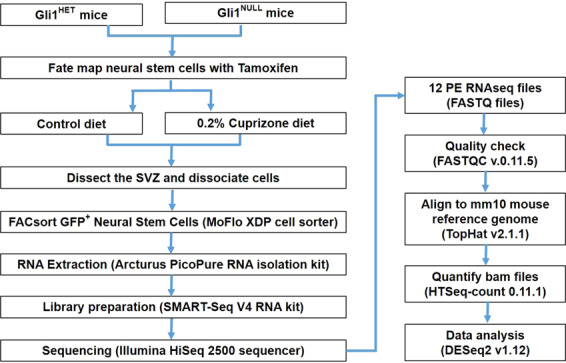
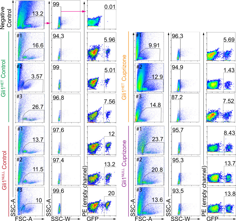
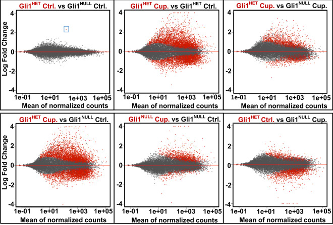
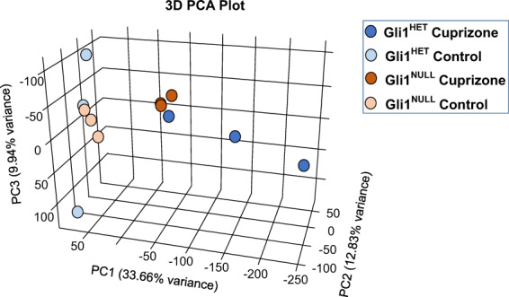
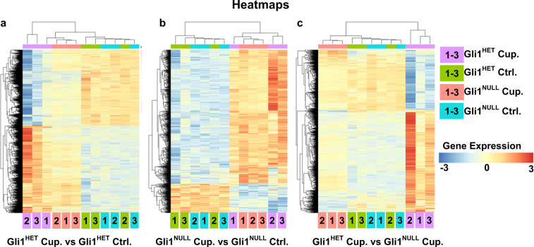
In the adult mammalian brain, Gli1 expressing neural stem cells reside in the subventricular zone and their progeny are recruited to sites of demyelination in the white matter where they generate new oligodendrocytes, the myelin forming cells. Remarkably, genetic loss or pharmacologic inhibition of Gli1 enhances the efficacy of remyelination by these neural stem cells. To understand the molecular mechanisms involved, we performed a transcriptomic analysis of this Gli1-pool of neural stem cells. We compared murine NSCs with either intact or deficient Gli1 expression from adult mice on a control diet or on a cuprizone diet which induces widespread demyelination. These data will be a valuable resource for identifying therapeutic targets for enhancing remyelination in demyelinating diseases like multiple sclerosis.


Conflict of interest statement

A patent on the method of targeting GLI1 as a strategy to promote remyelination has been awarded, with J. L. Salzer, J. Samanta and G. Fishell listed as co-inventors. JLS is a consultant for and has ownership interests in Glixogen Therapeutics.

Figures

Fig. 1
Fig. 1
Schematic overview of the study design. The flowchart demonstrates the experimental design and data analysis.
Fig. 2
Fig. 2
Flow cytometry analysis displaying the gating strategy used to sort fate-mapped neural stem cells. The negative control (top row) displays the gating strategy highlighted by the magenta arrows. The same strategy was applied for all samples in the figure. Fate-mapped, (i.e. GFP+) neural stem cells were purified from each of the samples depicted in the figure and processed for RNAseq. Percentage of gated cells is displayed above the selected area and the sample number is indicated in the top left corner of each dot plot. SSC- Side scatter, FSC- Forward scatter.
Fig. 3
Fig. 3
Visualization of differential gene expression in Gli1Het and Gli1Null NSCs. MA plots of the RNAseq data show the differences in expression of genes between the two genotypes on control and cuprizone diets. The blue box highlights the only gene differentially expressed between Gli1Het and Gli1Null NSCs in the control diet groups, corresponding to Gli1 itself. Ctrl.- Control diet group, Cup.-Cuprizone diet group.
Fig. 4
Fig. 4
Principle component analysis (PCA) of the sequencing data. A 3D PCA plot shows segregation of the control diet samples from the cuprizone diet samples. The variance is higher in the Gli1Het samples compared to the Gli1Null samples.
Fig. 5
Fig. 5
Heatmaps of the differentially expressed genes (FDR < 0.05). (a) Comparison of Gli1Het NSCs following demyelination with cuprizone diet (Cup.) (purple) vs. healthy NSCs on control diet (Ctrl.) (green). (b) Comparison of Gli1Null NSCs following demyelination with cuprizone diet (Cup.) (red) vs. healthy NSCs on control diet (Ctrl.) (blue). (c) Comparison of the Gli1Het vs. Gli1Null NSCs following demyelination with cuprizone diet (Cup). The sample numbers are mentioned inside the color coded box for each condition, at the bottom of the heatmap. The gene expression levels are color coded with the highest upregulated genes in red and the most down-regulated genes in blue.

References

    1. Compston A, Coles A. Multiple sclerosis. Lancet. 2008;372:1502–1517. doi: 10.1016/S0140-6736(08)61620-7. - DOI - PubMed
    1. Smith EJ, Blakemore WF, McDonald WI. Central remyelination restores secure conduction. Nature. 1979;280:395–396. doi: 10.1038/280395a0. - DOI - PubMed
    1. Dubois-Dalcq M, Ffrench-Constant C, Franklin RJ. Enhancing central nervous system remyelination in multiple sclerosis. Neuron. 2005;48:9–12. doi: 10.1016/j.neuron.2005.09.004. - DOI - PubMed
    1. Trapp BD, Nave KA. Multiple sclerosis: an immune or neurodegenerative disorder? Annu Rev Neurosci. 2008;31:247–269. doi: 10.1146/annurev.neuro.30.051606.094313. - DOI - PubMed
    1. Bunge MB, Bunge RP, Ris H. Ultrastructural study of remyelination in an experimental lesion in adult cat spinal cord. J Biophys Biochem Cytol. 1961;10:67–94. doi: 10.1083/jcb.10.1.67. - DOI - PMC - PubMed

Publication types