Review
. 2022 Feb;27(2):803-818.
doi: 10.1038/s41380-021-01361-1. Epub 2021 Oct 28.

Age-related immune alterations and cerebrovascular inflammation

Affiliations
Review

Age-related immune alterations and cerebrovascular inflammation

Carson E Finger et al. Mol Psychiatry. 2022 Feb.

Abstract

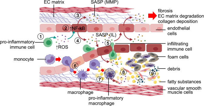
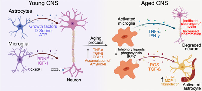
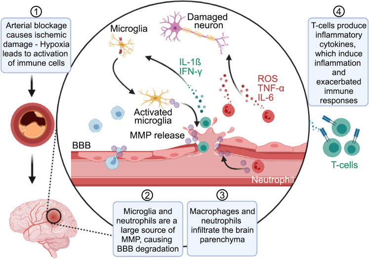
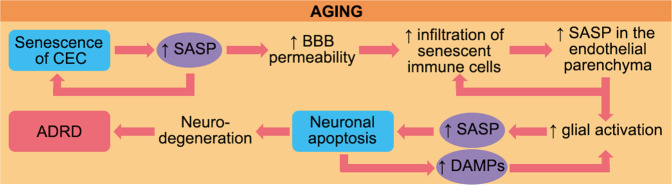
Aging is associated with chronic systemic inflammation, which contributes to the development of many age-related diseases, including vascular disease. The world's population is aging, leading to an increasing prevalence of both stroke and vascular dementia. The inflammatory response to ischemic stroke is critical to both stroke pathophysiology and recovery. Age is a predictor of poor outcomes after stroke. The immune response to stroke is altered in aged individuals, which contributes to the disparate outcomes between young and aged patients. In this review, we describe the current knowledge of the effects of aging on the immune system and the cerebral vasculature and how these changes alter the immune response to stroke and vascular dementia in animal and human studies. Potential implications of these age-related immune alterations on chronic inflammation in vascular disease outcome are highlighted.


Conflict of interest statement

The authors have no conflict of interest to disclose.

Figures

Fig. 1
Fig. 1. Age-related changes in the vasculature.
[1] Senescent immune cells secrete reactive oxygen species (ROS) that [2] activates the NF-kB pathway in cerebral endothelial cells (CECs). Then, CECs adopt a senescent-associated secretory phenotype (SASP) and [3] secrete MMP that degrade the extracellular matrix. Other SASP components secreted by senescent CECs can also promote fibrosis and collagen deposition. [4] Senescent CECs secrete pro-inflammatory substances (IL-1, IL-6, IL-8) into the vasculature lumen that impair tight junctions between CEC, and [5] facilitate the infiltration of immune cells and monocytes through the CEC layer. [6] Infiltrating monocytes reach the internal elastic lamina and change their phenotype to macrophages, [7] which phagocytize oxidized lipoproteins. In the internal elastic lamina, [8] reactive macrophages and infiltrating immune cells secrete pro-inflammatory cytokines that exacerbate inflammatory responses, and [9] contribute to the deposition of cellular debris, fatty substances, migrated vascular smooth muscle cells, and lipid-laden macrophages (foam cells) that lead to the formation of atherosclerotic plaques. Figure made with Biorender.com.
Fig. 2
Fig. 2. Astrocytes and microglia in a young CNS release growth factors and cellular signals to maintain homeostasis and control neurogenesis of neurons.
As individuals age, cellular and molecular changes in the brain environment are initiated by an increase of pro-inflammatory cytokines and an accumulation of proteins, such as amyloid. Next, microglia and the innate immune response are activated, activating astrocytes and leading to neuronal damage. Activation of microglia and astrocytes disrupts the BBB and contributes to a heightened immune response and worse cognitive outcomes in elderly patients with cerebrovascular injury. Neurological signals: ATP: adenosine triphosphate, BDNF: brain-derived neurotrophic factor, CX3CR1 or CX3CL1: fractalkine receptor and ligand IGF-1: Insulin-like growth factor, Aβ: amyloid-beta, IRF-7: Interferon regulatory factor 7, INF-γ: Interferon gamma, GFAP: Glial Fibrillary acidic protein, MCP-1: Macrophage chemoattractant protein, ROS: Reactive oxygen species, TGFβ, transforming growth factor-β, TNFα: tumor necrosis factor-alpha. Figure made with Biorender.com.
Fig. 3
Fig. 3. Cellular and molecular changes in the brain are initiated by primary brain injury.
In response to injury, damage-associated molecular patterns (DAMPs) are released, and an innate immune response characterized by glial activation and infiltration of blood-borne immune cells into the brain occurs. The activation and infiltration of peripheral immune cells lead to secondary brain injury, further destroying brain tissue and poor recovery. Figure made with Biorender.com.
Fig. 4
Fig. 4. After a brain insult, including ischemic stroke, activated microglia trigger an inflammatory response and eliminate debris from apoptotic cells.
After ischemic stroke, immune cells activate [1], and microglia secrete MMPs [2] that disrupt the integrity of the BBB and facilitates the invasion of macrophages and neutrophils into the brain parenchyma [3]. However, in the aged brain, this pro-inflammatory response is extended and contributes to the participation of T-cells that magnify the immune response [4]. Depletion of microglia prior to stroke exacerbated injury. One potential strategy to mitigate inflammation after brain injury is to deplete pathological microglia or enhance their capacity for repair. Figure made with Biorender.com.
Fig. 5
Fig. 5. Aging has detrimental effects in the regulation of important anti-inflammatory regulators associated with the cerebral vasculature.
The expression of ADAM metallopeptidase domain 10 (ADAM10), nuclear factor erythroid 2-related factor (Nrf2), and endothelial nitric oxidase synthase (eNOS) is downregulated with aging. ADAM10 cleavages amyloid-β precursor protein (APP) and forms soluble APPα, which opposite to soluble APPβ appears to be neuroprotective. Nrf2 negatively regulates the expression of β-secretase, which cleavages APP and form soluble APPβ. This contributes to the deposition of amyloid-β and promotes neuroinflammation. eNOS is synthesized by the cerebral endothelium and prevents oxidative stress. However, reduced levels of eNOS during aging enhances oxidative stress, which activates the TLR-NF-κB pathway axis and enhances the secretion of pro-inflammatory cytokines. In addition, the TLR-NF-κB pathway axis uncouples eNOS, creating a feedback loop that aggravates neuroinflammation. Blue indicates a beneficial effect for the cerebral vasculature and the brain, and red indicates a harmful effect.
Fig. 6
Fig. 6. Aging is the main risk factor for Alzheimer’s disease and related dementias (ADRD).
Aged individuals show an enhanced number of senescent cerebral endothelial cells (CECs) in their brains, which acquire a senescence-associated secretory phenotype (SASP) with detrimental consequences for tight junctions and the blood brain barrier (BBB) integrity. Thus, circulating senescent immune cells are more likely to penetrate between CECs and infiltrate into the endothelial parenchyma, where activated macrophages secrete pro-inflammatory cytokine. Glial cells also secrete molecules that contribute to the inflammation of the CNS and promote neuronal apoptosis. Further, danger-associated molecular patterns (DAMPs) derived from apoptotic neuron debris contribute to glial activation that sustains the CNS inflammation. Altogether, these events lead to neurodegeneration and contribute to ADRD and VCID. Note that SASP are “hot points” that exacerbate inflammation in the aged brain. Targeting the secretion of pro-inflammatory molecules from senescent cells and blocking the harmful feedback loops on CECs and neurons may prevent BBB disruption and neurodegeneration and thus counteract age related brain changes.

References

    1. Malaguarnera L, Ferlito L, Imbesi RM, Gulizia GS, Di Mauro S, Maugeri D, et al. Immunosenescence: a review. Arch Gerontol Geriatr. 2001;32:1–14. doi: 10.1016/S0167-4943(00)00086-8. - DOI - PubMed
    1. Nikolich-Žugich J. The twilight of immunity: emerging concepts in aging of the immune system. Nat Immunol. 2018;19:10–9. doi: 10.1038/s41590-017-0006-x. - DOI - PubMed
    1. Collaborators GBDLRoS. Feigin VL, Nguyen G, et al. Global, regional, and country-specific lifetime risks of stroke, 1990 and 2016. N Engl J Med. 2018;379:2429–37. doi: 10.1056/NEJMoa1804492. - DOI - PMC - PubMed
    1. Qu J, Chen Y, Luo G, Zhong H, Xiao W, Yin H. Delirium in the acute phase of ischemic stroke: incidence, risk factors, and effects on functional outcome. J Stroke Cerebrovasc Dis. 2018;27:2641–7. doi: 10.1016/j.jstrokecerebrovasdis.2018.05.034. - DOI - PubMed
    1. Moulin S, Leys D. Stroke occurring in patients with cognitive impairment or dementia. Arq Neuropsiquiatr. 2017;75:117–21. doi: 10.1590/0004-282x20160187. - DOI - PubMed

Publication types