Review
. 2021 Dec;22(12):741-757.
doi: 10.1038/s41583-021-00528-7. Epub 2021 Oct 28.

The neural mechanisms of manual dexterity

Affiliations
Review

The neural mechanisms of manual dexterity

Anton R Sobinov et al. Nat Rev Neurosci. 2021 Dec.

Abstract

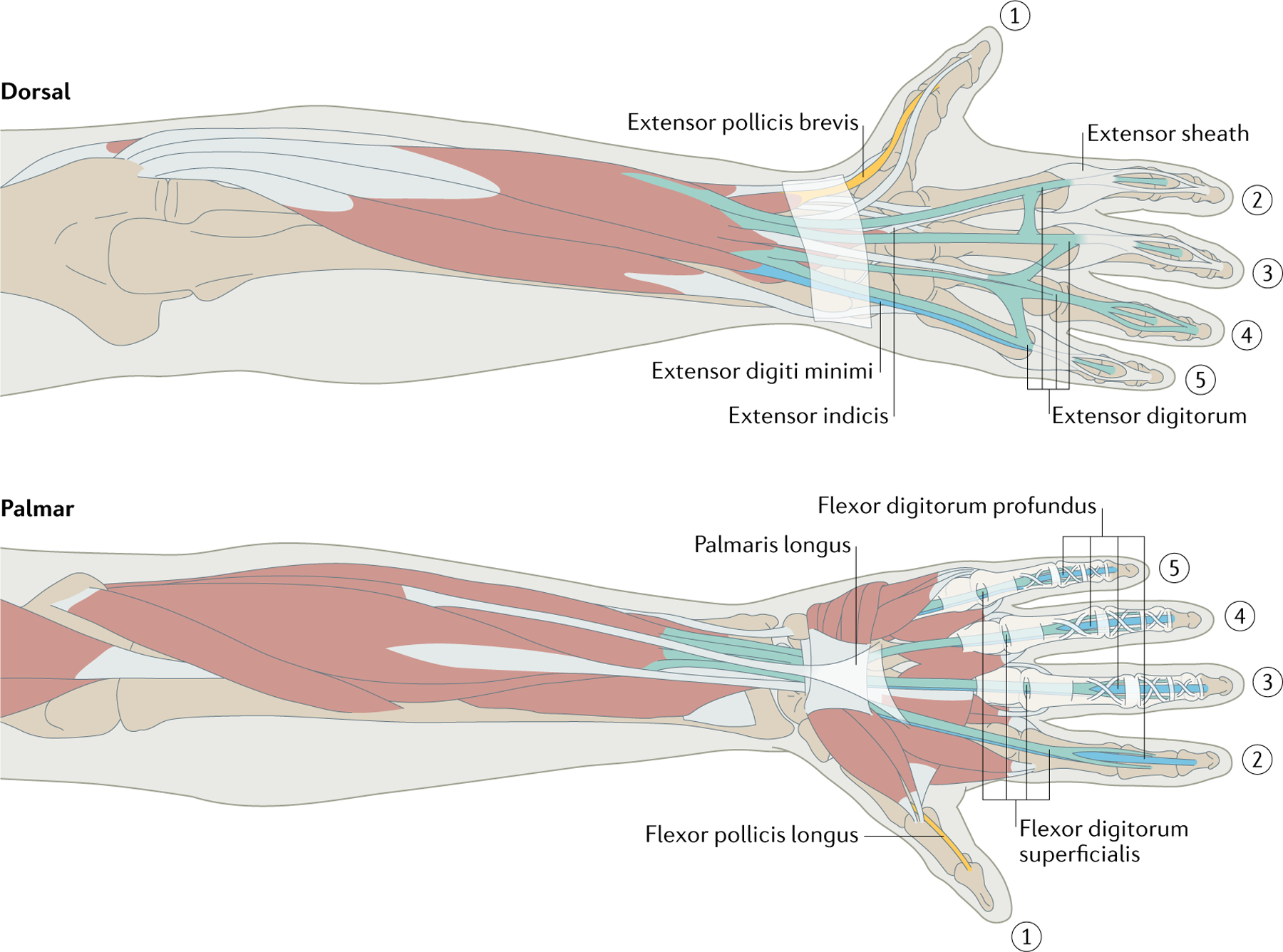
The hand endows us with unparalleled precision and versatility in our interactions with objects, from mundane activities such as grasping to extraordinary ones such as virtuoso pianism. The complex anatomy of the human hand combined with expansive and specialized neuronal control circuits allows a wide range of precise manual behaviours. To support these behaviours, an exquisite sensory apparatus, spanning the modalities of touch and proprioception, conveys detailed and timely information about our interactions with objects and about the objects themselves. The study of manual dexterity provides a unique lens into the sensorimotor mechanisms that endow the nervous system with the ability to flexibly generate complex behaviour.


Conflict of interest statement

Competing interests

The authors declare no competing interests.

Figures

Fig. 1 |
Fig. 1 |. Hand musculature.
Digits 2–5 are articulated mainly by two flexors — note how the deep one (blue) threads through the superficial one (green) at the proximal phalanx — and one extensor, which feeds into the extensor sheath. Intrinsic hand muscles also feed into the sheath, flexing the proximal finger joint and extending the other. Extensor indicis and extensor digiti minimi contribute further independence to index finger and little finger extension. Flexor pollicis longus and extensor pollicis brevis are two thumb muscles that are found only in humans and two other primate species. Palmaris longus lies outside the carpal tunnel and is absent in many people. Each muscle path is complex, especially around the wrist and thumb, wrapping around other moving muscles and bones, so estimating the action of these muscles is difficult. Some hand muscles and other tissues are omitted for clarity. In addition, the extensor sheath is omitted on the dorsal view of digit 4 and the connective tissue is omitted on the palmar view of digit 2 to reveal the underlying tendon paths. Image courtesy of Kenzie Green.
Fig. 2 |
Fig. 2 |. Direct and indirect pathways from the cortex to the muscles.
Traced axons (dots) from the primary motor cortex at the C8 level of the spinal cord. a | Such tracing has revealed that, in squirrel monkeys, neurons from the primary motor cortex (M1) project via the pyramidal tracts (blue region) to spinal interneurons (located in the yellow region), which in turn project to motor neurons (located in the red region). b | In capuchins, M1 sends direct projections to motor neurons in addition to indirect projections through spinal interneurons. Presumably owing in part to this direct pathway, capuchins are more dexterous than squirrel monkeys. CM, corticomotoneuronal. Adapted with permission from REF., Copyright 1993 Society for Neuroscience.
Fig. 3 |
Fig. 3 |. Main cortical regions and pathways involved in visuomotor control of the hand.
Visual information from the primary visual cortex (V1) is transformed in the parietal reach region (PRR) to guide reaching movements via the dorsal premotor area (PMd). Similarly, the anterior interparietal area (AIP) processes visual information about object shape to guide grasping movements via the ventral premotor area (PMv). The PMv and the PMd both project to the primary motor cortex (M1).
Fig. 4 |
Fig. 4 |. Body maps in the somatosensory cortex of a monkey.
Touch applied to a location on the body activates a spatially restricted population of neurons in the somatosensory cortex. Nearby neurons are activated by nearby patches of skin, leading to highly structured maps of the body, termed the ‘somatosensory homunculus’. In primates, the volume of the cortex devoted to the hand is very large. Within this volume, the neural representations of individual digits are spatially distinct. CS, central sulcus; D, digit; PCS, postcentral sulcus. Adapted with permission from REF., Wiley.
Fig. 5 |
Fig. 5 |. Neural coding of touch.
a | Reconstructed response of a population of slowly adapting type 1 (SA1) fibres and rapidly adapting (RA) fibres when embossed letters are scanned across the fingertip. The spatial pattern of activation in SA1 fibres and to a lesser extent in RA fibres carries a faithful representation of the stimulus. b | Responses of a Pacinian corpuscle-associated fibre (middle) to 40 repeated presentations of two textures (left) scanned across the skin. As shown in the spectrogram of the response (right), Pacinian corpuscle-associated fibres produce highly repeatable spiking patterns that differ across textures. This temporal code conveys information about fine texture. Part a adapted with permission from REF., PNAS. Part b adapted with permission from REF., PNAS.

References

    1. Billard A & Kragic D Trends and challenges in robot manipulation. Science 364, eaat8414 (2019). - PubMed
    1. Mathis A et al. DeepLabCut: markerless pose estimation of user-defined body parts with deep learning. Nat. Neurosci 21, 1281–1289 (2018). - PubMed
    1. Graving JM et al. DeepPoseKit, a software toolkit for fast and robust animal pose estimation using deep learning. eLife 8, e47994 (2019). - PMC - PubMed
    1. Jeannerod M The Neural and Behavioural Organization of Goal-Directed Movements (Clarendon Press/Oxford University Press, 1988).
    1. Santello M, Flanders M & Soechting JF Patterns of hand motion during grasping and the influence of sensory guidance. J. Neurosci 22, 1426–1435 (2002). - PMC - PubMed

Publication types