

Helicobacter pylori in gastric cancer: Features of infection and their correlations with long-term results of treatment
- PMID: 34712033
- PMCID: PMC8515796
- DOI: 10.3748/wjg.v27.i37.6290
Helicobacter pylori in gastric cancer: Features of infection and their correlations with long-term results of treatment
Abstract
Background: Helicobacter pylori (H. pylori) is a spiral-shaped bacterium responsible for the development of chronic gastritis, gastric ulcer, gastric cancer (GC), and MALT-lymphoma of the stomach. H. pylori can be present in the gastric mucosa (GM) in both spiral and coccoid forms. However, it is not known whether the severity of GM contamination by various vegetative forms of H. pylori is associated with clinical and morphological characteristics and long-term results of GC treatment.
Aim: To establish the features of H. pylori infection in patients with GC and their correlations with clinical and morphological characteristics of diseases and long-term results of treatment.
Methods: Of 109 patients with GC were included in a prospective cohort study. H. pylori in the GM and tumor was determined by rapid urease test and by immunohistochemically using the antibody to H. pylori. The results obtained were compared with the clinical and morphological characteristics and prognosis of GC. Statistical analysis was performed using the Statistica 10.0 software.
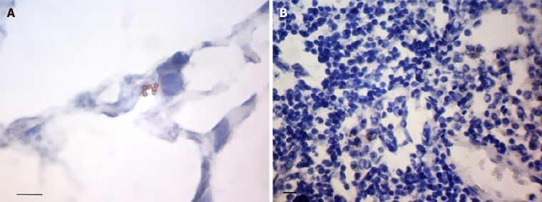
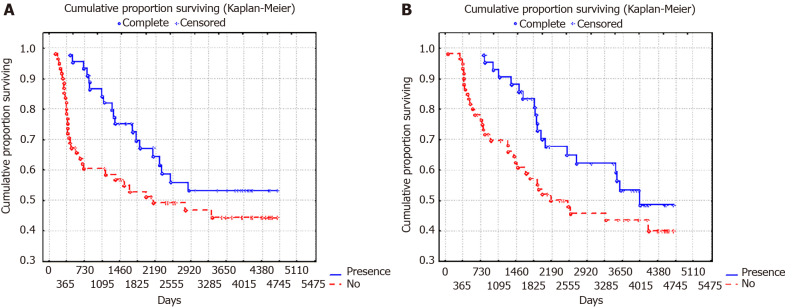
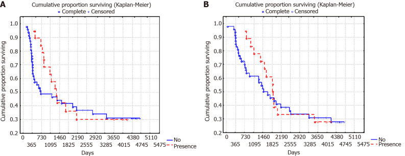
Results: H. pylori was detected in the adjacent to the tumor GM in 84.5% of cases, of which a high degree of contamination was noted in 50.4% of the samples. Coccoid forms of H. pylori were detected in 93.4% of infected patients, and only coccoid-in 68.9%. It was found that a high degree of GM contamination by the coccoid forms of H. pylori was observed significantly more often in diffuse type of GC (P = 0.024), in poorly differentiated GC (P = 0.011), in stage T3-4 (P = 0.04) and in N1 (P = 0.011). In cases of moderate and marked concentrations of H. pylori in GM, a decrease in 10-year relapse free and overall survival from 55.6% to 26.3% was observed (P = 0.02 and P = 0.07, respectively). The relationship between the severity of the GM contamination by the spiral-shaped forms of H. pylori and the clinical and morphological characteristics and prognosis of GC was not revealed.
Conclusion: The data obtained indicates that H. pylori may be associated not only with induction but also with the progression of GC.
Keywords: Coccoid and spiral forms of bacteria; Gastric cancer; Helicobacter pylori; Overall survival; Rapid urease test; Relapse free survival.
©The Author(s) 2021. Published by Baishideng Publishing Group Inc. All rights reserved.
Conflict of interest statement
Conflict-of-interest statement: The authors declare no conflicts of interests related to the publication of this study.
Figures


References
-
- Bray F, Ferlay J, Soerjomataram I, Siegel RL, Torre LA, Jemal A. Global cancer statistics 2018: GLOBOCAN estimates of incidence and mortality worldwide for 36 cancers in 185 countries. CA Cancer J Clin. 2018;68:394–424. - PubMed
-
- Siegel RL, Miller KD, Jemal A. Cancer statistics, 2015. CA Cancer J Clin. 2015;65:5–29. - PubMed
MeSH terms
LinkOut - more resources
Full Text Sources
Medical
Miscellaneous
