

Reconstruction of Complex Scalp Defects in Different Locations: Suggestions for Puzzle
- PMID: 34712077
- PMCID: PMC8526234
- DOI: 10.14744/SEMB.2020.98475
Reconstruction of Complex Scalp Defects in Different Locations: Suggestions for Puzzle
Abstract
Objective: Scalp defects may occur following trauma, radiotherapy, oncologic resection, and recurrent surgeries. The hair-bearing scalp has a dual role, which consists of protecting the calvarium and contributing to aesthetic appearance. While the "reconstructive ladder" approach may be used to close small and medium-sized scalp defects, it is not the case for larger ones involving the calvarium or with a radiation therapy history. The aim of this study is to present cases operated due to complex scalp defects, analyze complications, and discuss the choice of reconstruction.
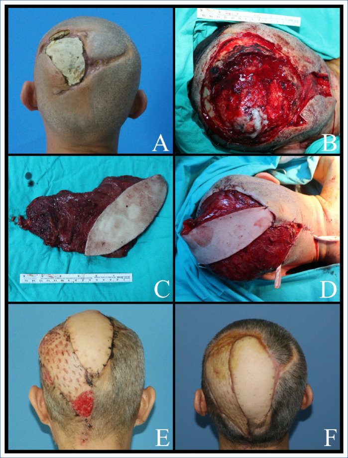
Material and methods: The study consists of 14 patients who were operated between December 2017 and August 2019 due to a complex scalp defect. Patient were evaluated according to age, gender, etiology, radiation therapy history, defect size and location, reconstruction steps, cranioplasty and duraplasty options, type of free flap, recipient artery, vein graft requirement, and complications.
Results: The mean age of patients, which consists of 11 men and three women, was 56.7 years. The etiology for scalp defects included basosquamous carcinoma, squamous cell carcinoma, giant basal cell carcinoma, atypical meningioma, glioblastoma multiforme, angiosarcoma, and anaplastic oligodendroglioma. The defect involved the full thickness of calvarium in nine cases and pericranium in five cases. Cranioplasties were made with rib graft (n=1), bone graft (n=1), and titanium mesh (n=7). Free flaps used for reconstruction were musculocutaneous latissimus dorsi (LD) (n=4), LD muscle (n=3), anterolateral thigh (ALT) (n=4), musculocutaneous ALT (n=1), vastus lateralis muscle (1), and rectus abdominis muscle (n=1). Flap loss was not observed. Complications occurred in four of the patients; include a partial graft loss, a wound dehiscence, seroma, and an unsatisfactory esthetic result.
Conclusion: Free tissue transfers rather than local flaps should be opted to reconstruct complex scalp defects, as failure of the latter, could create much greater defects, and worse consequences. There are many options for proper reconstruction, and it is essential to select the appropriate one, taking into account the comorbid conditions of each case.
Keywords: Free flaps; microsurgery; reconstruction; scalp.
Copyright: © 2021 by The Medical Bulletin of Sisli Etfal Hospital.
Conflict of interest statement
Conflict of Interest: None declared.
Figures


References
-
- Bradford BD, Lee JW. Reconstruction of the forehead and scalp. Facial Plast Surg Clin North Am. 2019;27:85–94. - PubMed
-
- Browne JD, Burke AJ. Benefits of routine maxillectomy and orbital reconstruction with the rectus abdominis free flap. Otolaryngol Head Neck Surg. 1999;121:203–9. - PubMed
-
- Chao AH, Yu P, Skoracki RJ, Demonte F, Hanasono MM. Microsurgical reconstruction of composite scalp and calvarial defects in patients with cancer:a 10-year experience. Head Neck. 2012;34:1759–64. - PubMed
-
- Chou PY, Lin CH, Hsu CC, Yang WH, Kane AA, Lin CH. Salvage of postcranioplasty implant exposure using free tissue transfer. Head Neck. 2017;39:1655–61. - PubMed
-
- Christy MR, Lipschitz A, Rodriguez E, Chopra K, Yuan N. Early postoperative outcomes associated with the anterolateral thigh flap in Gustilo IIIB fractures of the lower extremity. Ann Plast Surg. 2014;72:80–3. - PubMed
LinkOut - more resources
Full Text Sources
Research Materials