

Three cases of organized hematoma of the maxillary sinus in patients who underwent preoperative arterial embolization
- PMID: 34712374
- PMCID: PMC8529391
- DOI: 10.1016/j.radcr.2021.09.039
Three cases of organized hematoma of the maxillary sinus in patients who underwent preoperative arterial embolization
Erratum in
-
Erratum regarding missing patient consent statements in previously published articles.Radiol Case Rep. 2022 Nov 25;18(2):730-731. doi: 10.1016/j.radcr.2022.10.049. eCollection 2023 Feb. Radiol Case Rep. 2022. PMID: 36588598 Free PMC article.
Abstract
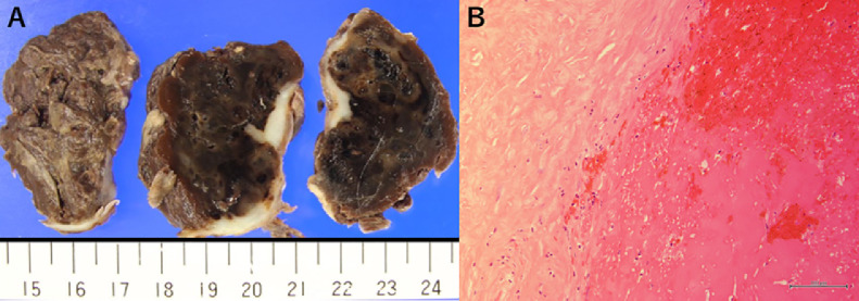
Organized hematoma (OH) is benign tumor in the maxillary sinus. The standard treatment for OH is complete surgical resection, however massive bleeding can occur during the procedure, albeit rarely. Some reports have suggested preoperative embolization is useful for reducing the volume of intraoperative bleeding. We report 3 cases of OH in the maxillary performed preoperative embolization. We identified the feeding arteries by angiography or IVR-CT, and we embolized them using Gelatin sponge particles. The embolized artery was the maxillary artery or both the maxillary and the facial artery. There were no major complications as a result of embolization. The mean fluoroscopy time was 35.8 minutes, and the mean fluoroscopy dose was 329.3 mGy. Tumor resection was performed the next day after arterial embolization. The mean bleeding volume for surgery was 383.3 ml, and the mean operative time was 194 minutes. No recurrence was observed in any of the cases over a 4-year follow-up period. We considered that it is possible that preoperative artery embolization is useful for decreasing intraoperative bleeding volume. Although the methods and usefulness of embolization await future reports, it is a technique that should be considered preoperatively because of its potential to prevent massive bleeding.
Keywords: Interventional Radiology; Intraoperative Bleeding; Maxillary Sinus; Organized Hematoma; Preoperative Embolization.
© 2021 The Authors. Published by Elsevier Inc. on behalf of University of Washington.
Figures


References
Publication types
LinkOut - more resources
Full Text Sources
