. 2021 Jun;4(6):510-522.
doi: 10.1038/s41929-021-00633-x. Epub 2021 Jun 21.

DYW domain structures imply an unusual regulation principle in plant organellar RNA editing catalysis

Affiliations

DYW domain structures imply an unusual regulation principle in plant organellar RNA editing catalysis

Mizuki Takenaka et al. Nat Catal. 2021 Jun.

Abstract

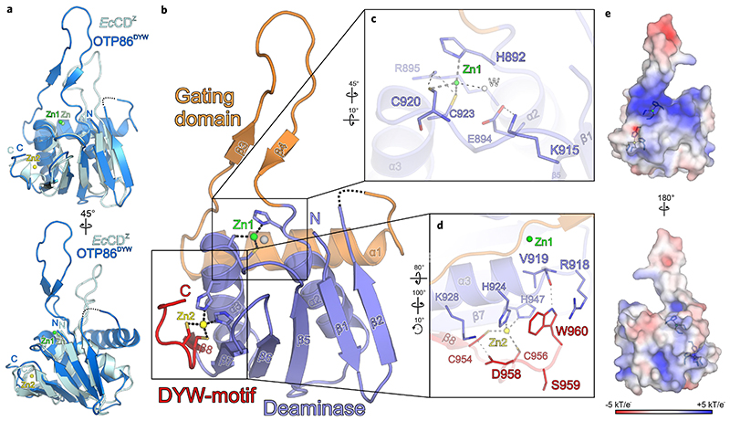
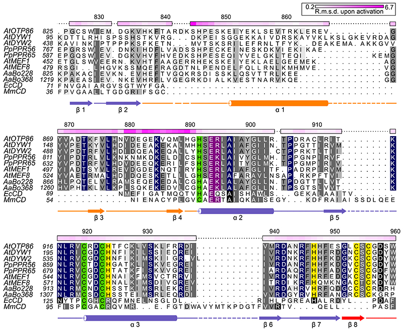
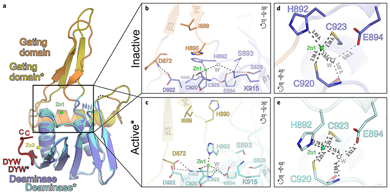
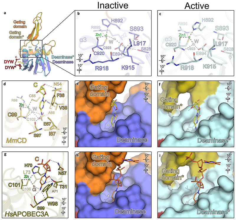
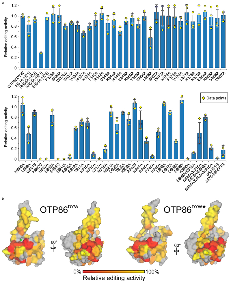
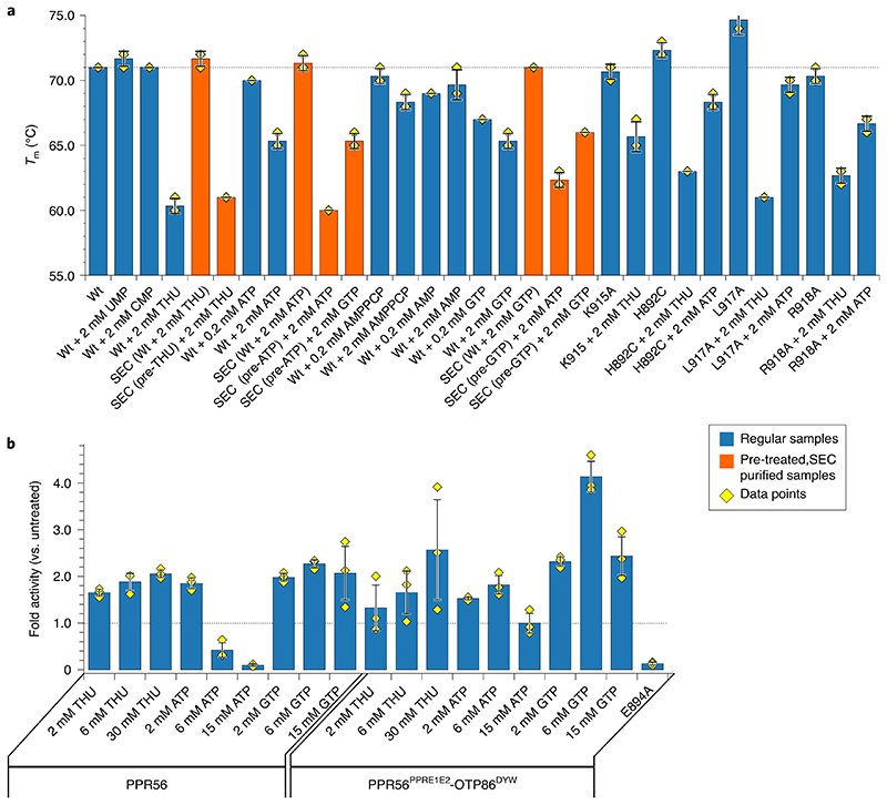
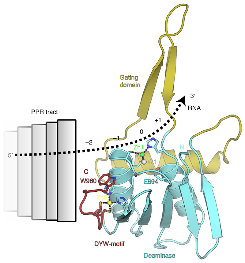
RNA editosomes selectively deaminate cytidines to uridines in plant organellar transcripts-mostly to restore protein functionality and consequently facilitate mitochondrial and chloroplast function. The RNA editosomal pentatricopeptide repeat proteins serve target RNA recognition, whereas the intensively studied DYW domain elicits catalysis. Here we present structures and functional data of a DYW domain in an inactive ground state and activated. DYW domains harbour a cytidine deaminase fold and a C-terminal DYW motif, with catalytic and structural zinc atoms, respectively. A conserved gating domain within the deaminase fold regulates the active site sterically and mechanistically in a process that we termed gated zinc shutter. Based on the structures, an autoinhibited ground state and its activation are cross-validated by RNA editing assays and differential scanning fluorimetry. We anticipate that, in vivo, the framework of an active plant RNA editosome triggers the release of DYW autoinhibition to ensure a controlled and coordinated cytidine deamination playing a key role in mitochondrial and chloroplast homeostasis.


Conflict of interest statement

Competing interests The authors declare no competing interests.

Figures

Fig. 1
Fig. 1. Crystal structure of the A. thaliana OTP86 DYW domain.
a, Superimposition of OTP86DYW (marine) with E. coli cytidine deaminase (EcCDZ, cyan) bound to the inhibitor zebularine (not shown) (PDB-ID: 1CTU; ref. ). The consensus deaminase zinc ions are shown as green (OTP86DYW, Zn1) and light-green (EcCDZ, Zn) spheres, a zinc ion partially coordinated by the DYW motif is shown in yellow (Zn2). b, The OTP86DYW structure defines a paradigmatic organization for DYW domains. The cytidine deaminase domain (slate) coordinates a zinc ion (Zn1, green) three-fold with H892, C920 and C923, the fourth position is occupied by a water molecule (W, white sphere). The deaminase domain is interrupted by a gating domain (orange) and terminates with a DYW motif (red), partially coordinating a second zinc ion (Zn2, yellow). c, A close-up view on the cytidine deaminase active site, with catalytically relevant residues shown as sticks. d, A close-up view of the DYW motif and the flanking β-strand 7 as well as α-helix 3. e, Electrostatic surface potentials as indicated by the colour scale bar (bottom), obtained by APBS version 1.5 and plotted on the surface of OTP86DYW. Residues involved in zinc coordination are shown as sticks; zinc atoms are as in b. Rotation symbols indicate the views relative to b. Interacting residues are shown as sticks and coloured by atom type. Blue, nitrogen; red, oxygen; yellow, sulfur; carbons take the colour of the respective molecule. Dashed lines represent hydrogen bonds, whereas thick grey dashed lines indicate zinc coordination. Dashed lines in the ribbon plots represent residues 842-844 not clearly defined by electron density.
Fig. 2
Fig. 2. Structure-based sequence alignment of OTP86DYW.
The alignment compares OTP86DYW with plant organellar DYW domains, hornwort putative U-to-C aminases,, C-to-U deaminases from E. coli (EcCD, PDB-ID: 1AF2; ref. ), and Mus musculus (MmCD, PDB-ID: 2FR6; ref. ). The alignment was prepared by Chimera employing Clustal Omega and shaded with ALSCRIPT,. Proteins are identified on the left of the aligned sequences with residues numbered (see Supplementary Table 2). Higher conservation is indicated by a darker background. Full conservation within DYW domains is indicated by dark blue background shading. Single-residue Cα r.m.s.d. values (calculated with CCP4i) between OTP86DYW and OTP86DYW (or structural changes upon activation) are displayed on the top of each alignment block as a colour gradient from white to pink (prepared with Python Matplotlib) and are quantified in the top-right box. The overall r.m.s.d. between the two structures was 1.99 Å, the gating domain residues 870–891 (β-finger) amounted to 2.98 Å. The numbering on top of each block refers to A. thaliana OTP86. Below the alignment blocks, secondary structure elements (αα-helix, β-β-sheet) of OTP86DYW are shown in slate for the deaminase domain, orange for the gating domain and red for the DYW motif. A dashed line or no line indicates residues missing from the structure or the expression construct, respectively. Green shading indicates residues coordinating the catalytic Zn1 ion; yellow shading indicates residues coordinating the Zn2 atom within the DYW motif and strand β7/helix α3; and purple shading indicates residues relevant for catalysis according to past studies. At, Arabidopsis thaliana; Pp, Physcomitrium patens; Aa, Anthoceros agrestis.
Fig. 3
Fig. 3. The DYW gating domain regulates cytidine deamination catalysis.
a, Superimposition of inactive OTP86DYW (colouring and dashed lines are as in Fig. 1b) and activated OTP86DYW (deaminase domain, cyan; gating domain, ochre; DYW motif, dark red). Zn1 (green) and Zn2 (yellow) of OTP86DYW are shown. b, A close-up view of the OTP86DYW active site in the inhibited state. c, A close-up view of the activated OTP86DYW active site in a catalytically competent conformation. d, A close-up view of the Zn1-coordination environment of the OTP86DYW active site in its catalytically inhibited state. e, A close-up view of the Zn1-coordination environment of the OTP86DYW active site in its catalytically active state. Distances within the vicinity of Zn1 are given in Å. Rotation symbols indicate the views relative to a. Interacting residues are shown as sticks and coloured by atom type. Water molecules (W) shown as white spheres. Carbon — as for the respective molecule; nitrogen, blue; oxygen, red; sulfur, yellow. Dashed lines represent hydrogen bonds, thick grey dashed lines indicate zinc coordination.
Fig. 4
Fig. 4. The OTP86DYW active site is sterically regulated by the gating domain.
a, Superimposition of inactive OTP86DYW and activated OTP86DYW (as in Fig. 3a). b, A close-up-view of the OTP86DYW active site in the inhibited state, depicting catalytic and potential RNA binding residues. c, A close-up-view of the OTP86DYW active site. d, M. musculus cytidine deaminase (MmCD; light orange) in complex with cytidine (yellow), coordinated zinc (green sphere) and an activated water molecule (white sphere) (PDB-ID: 2FR6; ref. ) e,f, A surface display of the active site cavity of OTP86DYW (e) and OTP86DYW (f) with the superimposed cytidine from d. g, Human APOBEC3A (dark ochre) with bound DNA (orange, only the active site cytidine is shown for clarity), coordinated Cl (light pink) (PDB-ID: 5KEG; ref. ). h,i, A surface display of the active site cavity of OTP86DYW (h) and of OTP86DYW (i) with the superimposed DNA from g. The cytidine deaminase structure of M. musculus and human APOBEC3A were superimposed employing only the zinc-coordinating residues and the equivalents of OTP86DYW E894. The colouring and dashes are as in Fig. 3. Rotation symbols indicate the views relative to a.
Fig. 5
Fig. 5. Orthogonal in vivo RNA editing validates the OTP86DYW domain structure and activation.
a, The C-to-U editing activities at the nad4eU272SL site in E. coli expressed with the PPR56PPRE1E2-OTP86DYW fusion protein (OTP86DYW), or its mutants, are plotted. The activities of mutants are relative to that of PPR56PPRE1E2-OTP86DYW (82.4 ± 2.1% edited). The bars represent the mean values, with each mutant protein ±s.d. based on three independent experiments (shown as yellow diamonds). The soluble protein expression of each mutated construct in E. coli was verified by western blot analysis (Supplementary Fig. 7). b, The activities of OTP86DYW mutants shown in a plotted on the surface of the inactive OTP86DYW and the activated OTP86DYW structure as a heatmap (activity is scaled in the bar on the bottom), with untested residues shown in grey.
Fig. 6
Fig. 6. in vitro editing and thermal shift assays validate the activation principle of OTP86DYW.
a, Melting points of OTP86DYW and selected variants in the presence of substrate, product and activators. Thermal shift assays were performed in triplicates. Curves were subjected to sigmoidal fitting; error bars indicate the s.d. of the three measurements (shown as yellow diamonds); regular samples are shown as blue bars, whereas SEC-related experiments are shown as orange bars; ‘pre’ indicates a sample pre-incubated with the respective activator and purified by SEC. b, C-to-U conversion for in vitro reactions with recombinant PPR56 or PPR56PPRE1E2-OTP86DYW with the addition of THU, ATP or GTP are displayed in a bar plot. In vitro editing with recombinant PPR56PPRE1E2-OTP86DYW-E894A mutant protein showed no editing activity. Bars indicate the mean value ±s.d. based on three independent experiments (shown as yellow diamonds). A grey dashed line indicates a T m of 71°C for wild type protein (WT in a) or the activity of the untreated proteins (in b).
Fig. 7
Fig. 7. Model for the anticipated RNA path across OTP86DYW in an RNA editosome context.
Activation of OTP86DYW is triggered by a conformational change of its gating domain, which may be elicited by an activator such as ATP, PPR–RNA complex formation, via the E1/E2 domains or different factors such as MORF proteins. The direction and path of the RNA can be extrapolated from comparisons to known cytidine deaminase co-structures such as human APOBEC3A (see Fig. 4h,i; PDB-ID: 5KEG), our comprehensive mutations of the domain surface (see Fig. 5) and the charged surface of OTP86DYW (see Fig. 1e). Colours and labels are as in Fig. 3a.

References

    1. Hiesel R, Wissinger B, Schuster W, Brennicke A. RNA editing in plant mitochondria. Science. 1989;246:1632–1634. - PubMed
    1. Covello PS, Gray MW. RNA editing in plant mitochondria. Nature. 1989;341:662–666. - PubMed
    1. Gualberto JM, Lamattina L, Bonnard G, Weil JH, Grienenberger JM. RNA editing in wheat mitochondria results in the conservation of protein sequences. Nature. 1989;341:660–662. - PubMed
    1. Hoch B, Maier RM, Appel K, Igloi GL, Kössel H. Editing of a chloroplast mRNA by creation of an initiation codon. Nature. 1991;353:178–180. - PubMed
    1. Takenaka M, Zehrmann A, Verbitskiy D, Härtel B, Brennicke A. RNA editing in plants and its evolution. Annu Rev Genet. 2013;47:335–352. - PubMed