. 2022 Apr;32(4):2639-2649.
doi: 10.1007/s00330-021-08317-7. Epub 2021 Oct 29.

COVID-19 pneumonia imaging follow-up: when and how? A proposition from ESTI and ESR

Affiliations

COVID-19 pneumonia imaging follow-up: when and how? A proposition from ESTI and ESR

K Martini et al. Eur Radiol. 2022 Apr.

Abstract

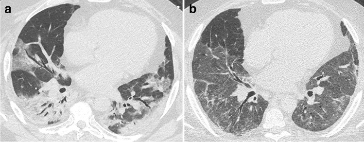
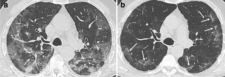
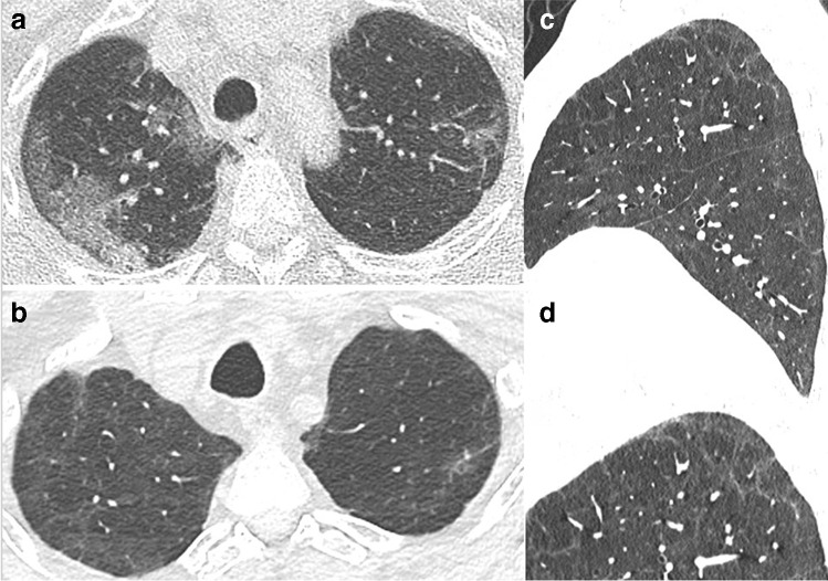
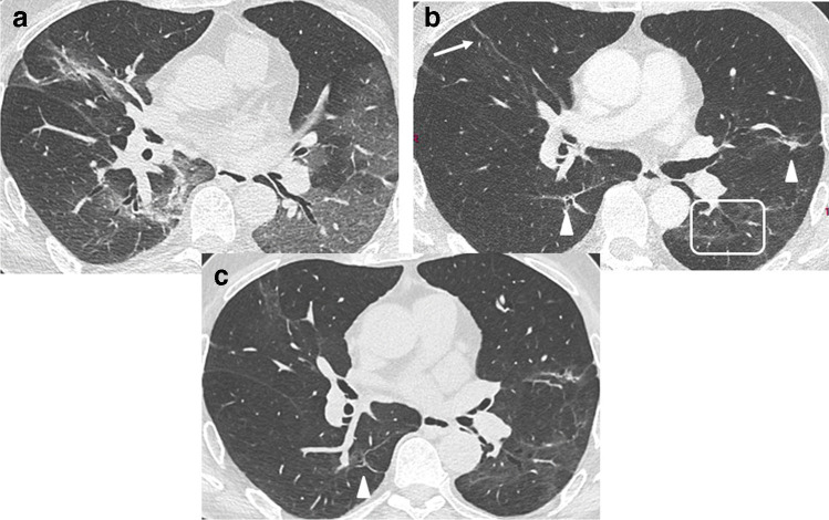
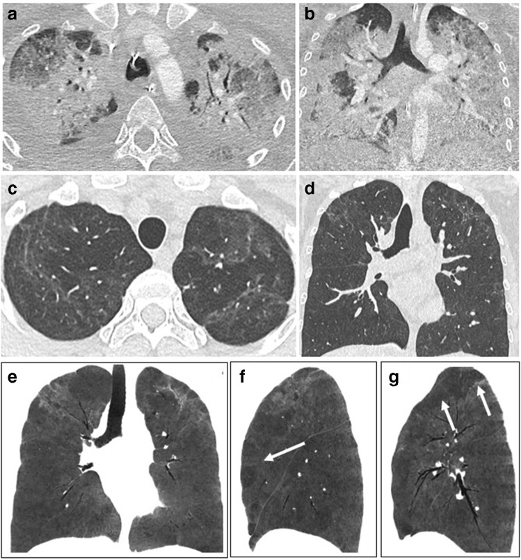
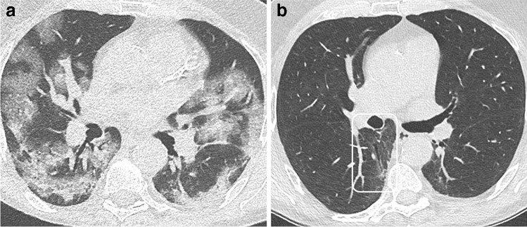
This document from the European Society of Thoracic Imaging (ESTI) and the European Society of Radiology (ESR) discusses the role of imaging in the long-term follow-up of COVID-19 patients, to define which patients may benefit from imaging, and what imaging modalities and protocols should be used. Insights into imaging features encountered on computed tomography (CT) scans and potential pitfalls are discussed and possible areas for future review and research are also included. KEY POINTS: • Post-COVID-19 pneumonia changes are mainly consistent with prior organizing pneumonia and are likely to disappear within 12 months of recovery from the acute infection in the majority of patients. • At present, with the longest series of follow-up examinations reported not exceeding 12 months, the development of persistent or progressive fibrosis in at least some individuals cannot yet be excluded. • Residual ground glass opacification may be associated with persisting bronchial dilatation and distortion, and might be termed "fibrotic-like changes" probably consistent with prior organizing pneumonia.

Keywords: COVID-19; Diagnostic imaging; Follow-up; Lung; Multidetector computed tomography.


Conflict of interest statement

The authors of this manuscript declare no relationships with any companies whose products or services may be related to the subject matter of the article.

Figures

Fig. 1
Fig. 1
CT features at baseline and at discharge in a 52-year-old man who developed severe COVID-19 pneumonia (intubation, extracorporeal membrane oxygenation, 45 days in ICU). a Baseline CT showing bilateral consolidation and ground glass opacities (GGOs) with peripheral and peribronchovascular distribution. b 2-month CT follow-up demonstrating the complete resolution of consolidation, with residual parenchymal bands bilaterally, while GGOs appear more extensive (“tinted sign”) in the lower lobes, lingula, and right middle lobe. Note the improved expansion of the left lung in b
Fig. 2
Fig. 2
CT features at baseline and 3-month follow-up, in a 60-year-old man with initial medium severity (10-day oxygen supplementation). a Baseline CT showing bilateral peripheral ground glass opacities (GGOs). b 3-month CT follow-up. GGOs significantly improved. Some areas of linear consolidation parallel to the pleura are demonstrated (arrows), consistent with late phase organizing pneumonia. Mild deformation of the left major fissure is noted, but there are no formal signs of fibrosis such as traction bronchiectasis or honeycombing
Fig. 3
Fig. 3
CT features at baseline, and 4-month follow-up, in a 52-year-old woman who developed severe COVID-19 pneumonia (intubation, 42 days in ICU). a Baseline CT. b, c 4-month CT follow-up. Ground glass opacities (GGOs) have partially resolved (low-density GGO), with remaining linear opacities on the axial transverse view through the upper lung (b). On the sagittal reformation (c), the polygonal shape suggests that linear opacities on the axial transverse view have a perilobular distribution (d, magnification), which might indicate late phase organizing pneumonia
Fig. 4
Fig. 4
CT features at baseline and 4-month and 1-year follow-up, in a 65-year-old man who developed severe COVID-19 pneumonia (intubation, 27 days in ICU). a Baseline CT. b 4-month CT follow-up. Ground glass opacities (GGOs) have largely resolved, except in the upper segment of the left lower lobe. Bronchial dilatation (square) is demonstrated within the residual GGO area. Linear consolidation (arrow head) and parenchymal band (arrow) are also denoted. c 1-year follow-up. CT features are unchanged. There are no honeycombing and no peripheral reticulations
Fig. 5
Fig. 5
CT features at baseline, and 1-year follow-up, in a 30-year-old woman who developed severe COVID-19 pneumonia (intubation, 40 days in ICU). a, b Baseline CT showing extensive ground glass opacities (GGOs) and consolidation. cg 1-year CT follow-up. Residual GGO, with a linear shape, is mainly seen in the upper lung, suggestive of organizing pneumonia at a late phase. There is no bronchial dilatation, signs of architectural distortion, or honeycombing, as confirmed by minimum intensity projections (minIP) (e, f, g). The sagittal minIP reformations (f, g) demonstrate plurilobular areas of decreased attenuation (arrows). This mosaic attenuation pattern might be due to either residual small airway disease or peripheral vascular obstruction. DLCO at 1 year was 64%
Fig. 6
Fig. 6
CT features at baseline, and 1-year follow-up, in a 63-year-old man who developed severe COVID-19 pneumonia (intubation, 9 days in ICU). a Baseline CT. b 1-year CT follow-up. Ground glass opacities (GGOs) have largely resolved. Mild bronchial dilatation (rectangle) is demonstrated within the residual GGO area of the right lower lobe. Pulmonary function tests are normal, with a DLCO value of 90%

References

    1. Mahase E. Covid-19: what do we know about “long covid”? BMJ. 2020;370:m2815. doi: 10.1136/bmj.m2815. - DOI - PubMed
    1. Greenhalgh T, Knight M, A’Court C, et al. Management of post-acute covid-19 in primary care. BMJ. 2020;370:m3026. doi: 10.1136/bmj.m3026. - DOI - PubMed
    1. Sudre CH, Murray B, Varsavsky T, et al. Attributes and predictors of long COVID. Nat Med. 2021;27:626–631. doi: 10.1038/s41591-021-01292-y. - DOI - PMC - PubMed
    1. Ojo AS, Balogun SA, Williams OT, Ojo OS. Pulmonary fibrosis in COVID-19 survivors: predictive factors and risk reduction strategies. Pulm Med. 2020;2020:e6175964. doi: 10.1155/2020/6175964. - DOI - PMC - PubMed
    1. Wunsch H. Mechanical Ventilation in COVID-19: interpreting the current epidemiology. Am J Respir Crit Care Med. 2020;202:1–4. doi: 10.1164/rccm.202004-1385ED. - DOI - PMC - PubMed