. 2021 Oct;10(5):e1237.
doi: 10.1002/mbo3.1237.

Polyunsaturated fatty acids cause physiological and behavioral changes in Vibrio alginolyticus and Vibrio fischeri

Affiliations

Polyunsaturated fatty acids cause physiological and behavioral changes in Vibrio alginolyticus and Vibrio fischeri

David S Smith et al. Microbiologyopen. 2021 Oct.

Abstract

Vibrio alginolyticus and Vibrio (Aliivibrio) fischeri are Gram-negative bacteria found globally in marine environments. During the past decade, studies have shown that certain Gram-negative bacteria, including Vibrio species (cholerae, parahaemolyticus, and vulnificus) are capable of using exogenous polyunsaturated fatty acids (PUFAs) to modify the phospholipids of their membrane. Moreover, exposure to exogenous PUFAs has been shown to affect certain phenotypes that are important factors of virulence. The purpose of this study was to investigate whether V. alginolyticus and V. fischeri are capable of responding to exogenous PUFAs by remodeling their membrane phospholipids and/or altering behaviors associated with virulence. Thin-layer chromatography (TLC) analyses and ultra-performance liquid chromatography-electrospray ionization mass spectrometry (UPLC/ESI-MS) confirmed incorporation of all PUFAs into membrane phosphatidylglycerol and phosphatidylethanolamine. Several growth phenotypes were identified when individual fatty acids were supplied in minimal media and as sole carbon sources. Interestingly, several PUFAs acids inhibited growth of V. fischeri. Significant alterations to membrane permeability were observed depending on fatty acid supplemented. Strikingly, arachidonic acid (20:4) reduced membrane permeability by approximately 35% in both V. alginolyticus and V. fischeri. Biofilm assays indicated that fatty acid influence was dependent on media composition and temperature. All fatty acids caused decreased swimming motility in V. alginolyticus, while only linoleic acid (18:2) significantly increased swimming motility in V. fischeri. In summary, exogenous fatty acids cause a variety of changes in V. alginolyticus and V. fischeri, thus adding these bacteria to a growing list of Gram-negatives that exhibit versatility in fatty acid utilization and highlighting the potential for environmental PUFAs to influence phenotypes associated with planktonic, beneficial, and pathogenic associations.

Keywords: Aliivibrio fischeri; Vibrio; Vibrio alginolyticus; biofilm; fatty acids; motility; phospholipids.


Conflict of interest statement

None declared.

Figures

FIGURE 1
FIGURE 1
Growth characteristics of Vibrio alginolyticus and Vibrio fischeri in the presence of exogenous polyunsaturated fatty acids. (a & b) V. fischeri was grown at 28℃ in CM9 minimal media (342 mM NaCl) with PUFAs as supplemental carbon sources [300 uM] (a) and as sole carbon sources [1mM] (b). (c & d) V. alginolyticus was grown at 28℃ in CM9 minimal media (342 mM NaCl) with PUFAs as supplemental carbon sources [300 uM] (c) and as sole carbon sources [1 mM] (d). (e & f) V. alginolyticus was grown at 37℃ in CM9 minimal media (150 mM NaCl) with PUFAs as supplemental carbon sources [300 uM] (e) and as sole carbon sources [1 mM] (f). Graphs are representative of at least two biological replicates (standard deviations <0.05).
FIGURE 2
FIGURE 2
Thin‐layer chromatography of phospholipids extracted from Vibrio alginolyticus and Vibrio fischeri grown in the presence of individual polyunsaturated fatty acids. Bacteria were grown to exponential phase (OD ≈ 0.8) in CM9 minimal media (3% NaCl) at 28℃ with or without 300 μM of the indicated fatty acids (linoleic acid [18:2], alpha‐linolenic acid [18:3α], gamma‐linolenic acid [18:3γ], dihomo‐gamma‐linolenic acid [20:3], arachidonic acid [20:4], eicosapentaenoic acid [20:5], and docosahexaenoic acid [22:6]). Phospholipids were extracted and subjected to separation by TLC (see Materials and Methods). Both V. alginolyticus (a) and V. fischeri (b) appear to be incorporating PUFAs into their phospholipid membranes. Compared to the “No FA” culture, the major bacterial phospholipids phosphatidylethanolamine (PE), phosphatidylglycerol (PG), and cardiolipin (CL) display a noticeable migratory shift upward. Linolenic acids (18:3α and 18:3γ) are not represented for V. fischeri because the bacteria do not grow to the logarithmic phase.
FIGURE 3
FIGURE 3
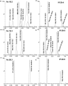
Ultra‐performance liquid chromatography/mass spectrometry of extracted lipids from Vibrio alginolyticus and Vibrio fischeri grown in the presence of a specific fatty acid. The phospholipid region of the chromatograms shows a variety of peaks corresponding to modified phospholipids that are absent in the control. Shown are extracted ion chromatograms for V. alginolyticus cultures exposed to (a) 18:2 (b) 18:3α (c) 20:3 and V. fischeri cultures exposed to (d) 20:4 (e) 20:5 (f) 22:6. Identifications of each phospholipid were based on mass spectrometry. See Appendix Figure A1 for assignment rationale.
FIGURE 4
FIGURE 4
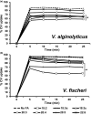
Effect of exogenous fatty acids on permeability in Vibrio alginolyticus and Vibrio fischeri. (a) V. alginolyticus and (a) V. fischeri were grown at 28℃ in CM9 (3% NaCl) with and without 300 µM of each PUFA to the mid‐log phase (OD = 0.8). Cultures were gently pelleted, washed, and prepared in PBS (OD600 = 0.7). Measurement of CV remaining in the supernatant at each time interval allowed calculation of the percentage of CV uptake. The graph is representative of three independent experiments. Standard deviations (not graphed for visual clarity) were less than 3% and statistical significance was measured by using all five‐time interval values compared to control (Student's t‐test, paired, 2‐tailed, *< 0.002).
FIGURE 5
FIGURE 5
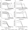
Effect of exogenous fatty acids on polymyxin B, colistin, and imipenem resistance in Vibrio alginolyticus and Vibrio fischeri. Bacteria were grown at 28℃ in CM9 (3% NaCl) with and without 300 µM of the indicated fatty acids to the mid‐log phase (OD = 0.8). Cultures were pelleted, washed, and resuspended in CM9 to a final desired inoculum of 5 × 105 cfu ml−1. Fatty acids were added to a final concentration of 300 μM. The bacterial suspension was transferred to microtiter plates containing twofold concentrations of (a & b) polymyxin B, (c & d) colistin, or (e & f) imipenem. Growth measurements (OD = 600nm) were recorded after 24 h incubation at 28℃. Experiments were performed in triplicate. Circled symbols indicate significant differences (< 0.002) as compared to the control (no fatty acid) at the same antimicrobial concentration.
FIGURE 6
FIGURE 6
Incubation with exogenous fatty acids alters biofilm formation in Vibrio alginolyticus and Vibrio fischeri. Overnight cultures were pelleted, washed, resuspended in the appropriate media, and inoculated onto microtiter plates (starting OD ~0.1) in octuplet. Each culture was grown in the presence of 300 μM of the indicated fatty acids. The biofilm assay was performed with V. alginolyticus (a & c) and V. fischeri (b & d) in CM9 minimal media (a & b) and Marine broth (c & d). Following incubation, the solubilized CV was measured at 590 nm. At least two independent biological replicates were performed in octuplets. The Student's t‐test was used to determine significant differences in biofilm formation (*,< 0.001).
FIGURE 7
FIGURE 7
Effects of PUFAs on swimming motility in Vibrio alginolyticus and Vibrio fischeri. For V. alginolyticus, soft agar motility plates were prepared with 10 g L−1 tryptone, 10 g L−1 NaCl, 0.35% agar, and with or without 300 μM of a given PUFA. For V. fischeri, soft agar motility plates were prepared using Marine broth containing 0.35% agar, and with or without 300 μM of a given PUFA. Overnight cultures were washed and resuspended to prepare a culture with an OD600 of 1.0. Each motility plate was divided into 4 quadrants and 2 µL of the diluted culture was injected into the center of each quadrant. (a) V. alginolyticus swimming motility was measured after 48, 72, and 96 h at 28℃. All fatty acids caused a steady decrease in swimming motility. (b) V. fischeri swimming motility was measured after 6h at 28℃. Asterisks indicate significant (< 0.01) deviations from the control sample at that incubation time, as calculated using a Student's t‐test (paired, two‐tailed distribution).
FIGURE A1
FIGURE A1
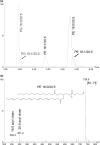
Ultra‐performance liquid chromatography/mass spectrometry of lipids isolated from Vibrio alginolyticus grown in the presence of eicosapentaenoic acid. Vibrio alginolyticus was grown to logarithmic phase at 28℃ in CM9 (3% NaCl) spiked with 300 μM of eicosapentaenoic acid (20:5). Electrospray ionization‐quadrupole mass spectrometry was used to detect [M‐H] ions produced following gradient elution using a reversed‐phase C18 column. The use of elevated cone voltage (50 V) produces cone fragments due to cleavage of the fatty acyl chains from the sn‐1 and sn‐2 positions. (a) Extracted ion chromatograms (overlain), mass filtered for the parent ions of the indicated phospholipids. These ions are absent in the control culture. (b) The mass spectrum of the chromatographic peak at 5.75 min displays a mass peak of 736.5 m/z which corresponds to a PE 36:5 species (http://www.lipidmaps.org/). Direct observation of the cone fragments at m/z 255.2 and 301.2 confirm the identity as PE 16:0/20:5. All chromatographic peaks were assigned in this way; by analysis of mass spectral parent peaks and their corresponding acyl chain, cone fragments were observable when using high cone voltage in negative mode electrospray ionization.
FIGURE A2
FIGURE A2
PUFAs increase biofilm formation of V. alginolyticus grown at human physiological temperature (37℃) and salt concentration (150mM NaCl). Overnight cultures were used to prepare fresh inocula in CM9 media (0.4% casamino acids, 0.4% glucose, 150 mM NaCl) and transferred onto microtiter plates (starting OD ~0.1) in octuplet. Growth conditions involved administration of 300μM of the indicated fatty acids and incubation at 37℃ for 48h. Absorbance values (OD590) were measured and data were expressed as the mean (± SD) of two independent experiments performed in octuplet. Asterisks indicate significant differences (< 0.001) in biofilm formation compared to control as determined by Student's t‐test.

Similar articles

Cited by

References

    1. Adin, D. M. , Phillips, N. J. , Gibson, B. W. , Apicella, M. A. , Ruby, E. G. , McFall‐Ngai, M. J. , Hall, D. B. , & Stabb, E. V. (2007). Characterization of htrB and msbB mutants of the light organ symbiont Vibrio fischeri . Applied and Environmental Microbiology, 74, 633–644. 10.1128/AEM.02138-07. - DOI - PMC - PubMed
    1. Ahmed, R. , Rafiquzaman, S. M. , Hossain, M. T. , Lee, J. M. , & Kong, I. S. (2015). Species‐specific detection of Vibrio alginolyticus in shellfish and shrimp by real‐time PCR using the groEL gene . Aquaculture International, 24, 157–170. 10.1007/s10499-015-9916-5. - DOI
    1. Ansari, M. N. A. , Nicolaides, N. , & Fu, H. C. (1970). Fatty acid composition of the living layer and stratum corneum lipids of human sole skin epidermis. Lipids, 5, 838–845. 10.1007/BF02531977. - DOI - PubMed
    1. Baker, L. Y. , Hobby, C. R. , Siv, A. W. , Bible, W. C. , Glennon, M. S. , Anderson, D. M. , Symes, S. J. , & Giles, D. K. (2018). Pseudomonas aeruginosa responds to exogenous polyunsaturated fatty acids (PUFAs) by modifying phospholipid composition, membrane permeability, and phenotypes associated with virulence. BMC Microbiology, 18, 117. 10.1186/s12866-018-1259-8. - DOI - PMC - PubMed
    1. Baron, S. , Hadjadj, L. , Rolain, J. M. , & Olaitan, A. O. (2016). Molecular mechanisms of polymyxin resistance: knowns and unknowns. International Journal of Antimicrobial Agents, 48(6), 583–591. 10.1016/j.ijantimicag.2016.06.023. - DOI - PubMed

Publication types

MeSH terms

LinkOut - more resources