. 2021 Oct 29;7(44):eabj7398.
doi: 10.1126/sciadv.abj7398. Epub 2021 Oct 29.

HIV-1 propagation is highly dependent on basal levels of the restriction factor BST2

Affiliations

HIV-1 propagation is highly dependent on basal levels of the restriction factor BST2

Balaji Olety et al. Sci Adv. .

Abstract

BST2 is an interferon-inducible antiviral host protein antagonized by HIV-1 Vpu that entraps nascent HIV-1 virions on the cell surface. Unexpectedly, we find that HIV-1 lacking Nef can revert to full replication competence simply by losing the ability to antagonize BST2. Using gene editing together with cell sorting, we demonstrate that even the propagation of wild-type HIV-1 is strikingly dependent on BST2, including in primary human cells. HIV-1 propagation in BST2−/− populations can be fully rescued by exogenous BST2 irrespective of its capacity to signal and even by an artificial BST2-like protein that shares its virion entrapment activity but lacks sequence homology. Counterintuitively, our results reveal that HIV-1 propagation is critically dependent on basal levels of virion tethering by a key component of innate antiviral immunity.


Figures

Fig. 1.
Fig. 1.. Rescue of Nef-deficient HIV-1 through loss of Vpu.
(A) Western blots showing that Nef-deficient HIV-1 gained the ability to propagate efficiently in MOLT-3 cells after several passages. The cells were infected with equal amounts (0.2 ng/ml p24) of Nef+ or Nef HIV-1NL4-3 freshly produced in 293T cells or of Nef HIV-1NL4-3 that had been passaged in MOLT-3 SERINC3/5 knockout cells. Cell lysates were examined with anti-capsid (CA) monoclonal antibody (mAb) and anti-Nef antiserum 8 days after infection. (B) Western blots showing that vpu and env sequences from the passaged virus allow Nef HIV-1NL4-3 to replicate vigorously in MOLT-3 cells. (C and D) The Nef/R1 recombinant, which harbors the disrupted vpu gene of the passaged virus, but not the Nef/R2 recombinant, which harbors its mutant env gene, replicates, as well as Nef+ HIV-1NL4-3 in MOLT-3 (C) and Jurkat E6.1 cells (D). Virus propagation was monitored by measuring p24 in the culture supernatants and by Western blotting of cell lysates. (E) A disrupted vpu initiation codon allows the naturally Nef-deficient HXBH10 strain to propagate in MOLT-3 cells, which are nonpermissive for Vpu+ HXBH10. (F) The Vpu version of HXBH10 also replicates with moderately accelerated kinetics in primary human cells. PBMC, peripheral blood mononuclear cells.
Fig. 2.
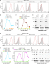
Fig. 2.. HIV-1 propagation is highly dependent on the Vpu target BST2.
(A) Expression of BST2 on parental MOLT-3 cells and on FACS-sorted subpopulations. The BST2+/+ (TS1) and BST2−/− (TS1) subpopulations were obtained by sorting after one round of gene editing with sgRNA TS1, and the BST2−/− (TS1/2) subpopulation was obtained by subjecting BST2−/− (TS1) cells to a second round of editing with sgRNA TS2, followed by a second round of sorting. Open histograms represent staining with anti-BST2 mAb; gray-shaded histograms represent the isotype control. (B and C) Virus growth curves showing that the propagation of WT (Vpu+/Nef+) HIV-1NL4-3 in the BST2−/− (TS1) subpopulation is highly impaired (B) and is even more impaired in the twice-sorted BST2−/− (TS1/2) subpopulation (C), which lacks detectable surface BST2. All subpopulations were infected with 0.2 ng p24/ml, and virus release was monitored by p24 enzyme-linked immunosorbent assay (ELISA). (D) Virus propagation in the same cultures monitored by comparing Gag expression levels in the infected cells at different days after infection by Western blotting with anti-CA. pI, post infection. (E) Expression of BST2 on FACS-sorted Jurkat E6.1 subpopulations. The BST2+/+ (TS1) and BST2−/− (TS1) subpopulations were obtained after gene editing with an sgRNA (TS1) that targets the BST2 gene, whereas two sgRNAs (TS3 and TS4) that target sites flanking the BST2 gene were used simultaneously to generate the BST2+/+ (TS3+4) and BST2−/− (TS3+4) subpopulations. (F) Propagation of WT HIV-1NL4-3 in Jurkat E6.1 subpopulations monitored by p24 ELISA after infection with 0.2 ng p24/ml. (G and H) Propagation of WT HIV-1NL4-3 in the BST2+/+ (TS3+4) and BST2−/− (TS3+4) subpopulations after infection with 0.2 or 2 ng p24/ml, monitored in parallel by p24 ELISA (G) and by Western blotting of cell lysates with anti-CA (H).
Fig. 3.
Fig. 3.. BST2 dependency is shared by R5-tropic HIV-1.
(A) CCR5 surface levels on parental MOLT-3 cells and on the twice-sorted BST2−/− (TS1/2) subpopulation after stable transduction with a retroviral vector expressing CCR5. (B) Replication of R5-tropic HIV-1 viruses in these cells monitored by comparing Gag expression levels by Western blotting with anti-CA after infection with 0.1 ng (NL-JRFL) or 0.2 ng (NL-ZM109) p24/ml. (C) Virus replication monitored in parallel by p24 ELISA.
Fig. 4.
Fig. 4.. BST2 is crucial for HIV-1 propagation in primary cells.
(A) BST2 levels on bulk PBMC subjected to gene editing with sgRNA targeting BST2 and on sorted subpopulations. The sorted cells were immediately reanalyzed by flow cytometry. The fraction of cells that fell within the gates set for sorting BST2−/− and BST2+/+ subpopulations is indicated by red and blue dashed lines, respectively. (B) Virus growth curves showing the replication of WT (Vpu+/Nef+) HIV-1NL4-3 in the sorted BST2+/+ and BST2−/− subpopulations after infection with 0.2 ng p24/ml in 1 ml of medium. (C) Replication of WT HIV-1NL4-3 in sorted BST2+/+ and BST2−/− PBMC subpopulations from additional three donors after infection with 0.5 ng p24/ml in 200 μl of medium. (D) Replication of an R5-tropic variant of WT HIV-1NL4-3 with a primary env gene in sorted BST2+/+ and BST2−/− PBMC subpopulations infected as in (C).
Fig. 5.
Fig. 5.. BST2 is dispensable for infectability but required for HIV-1 spreading.
(A) Dot plots showing that BST2+/+ and BST2−/− Jurkat E6.1/ZsGreen subpopulations are equally susceptible to single-cycle infection with cell-free WT (Vpu+/Nef+) HIV-1NL4-3. The entry inhibitor AMD3100 was added at 0 or 12 hours after infection with WT HIV-1NL4-3 at a high multiplicity of infection (MOI). Infected (ZsGreen+) cells were quantified by flow cytometry. (B) In the same cells, BST2 is essential for spreading infections. BST2+/+ and BST2−/− Jurkat E6.1/ZsGreen subpopulations were infected with a low amount of cell-free WT HIV-1NL4-3 (2 ng p24/ml), and after 10 days infected (ZsGreen+) cells were detected by fluorescence microscopy. FSC, forward scatter.
Fig. 6.
Fig. 6.. Innate sensing by BST2 is dispensable for its role in HIV-1 spreading.
(A) BST2 surface levels on Jurkat E6.1 BST2−/− knockout cells reconstituted with variants of human BST2. (B and C) Both signaling-competent and signaling-defective versions of human BST2 fully rescue WT (Vpu+/Nef+) HIV-1NL4-3 replication in the knockout cells, as examined by Western blotting of cell lysates with anti-CA on day 10 after infection with 0.2 ng p24/ml (B) and in parallel by p24 ELISA of culture supernatants (C). (D) BST2 surface levels on Jurkat E6.1 BST2−/− knockout cells reconstituted with mouse BST2. (E and F) Mouse BST2 partially restores WT HIV-1NL4-3 replication in the knockout cells, as examined by monitoring cell-associated Gag levels (E) and p24 antigen release (F). PE, phycoerythrin.
Fig. 7.
Fig. 7.. An artificial BST2-like molecule fully rescues HIV-1 propagation in BST2 knockout cells.
(A) Schematic illustration of an artificial BST2-like molecule (art-BST2) composed of sequences derived from the human transferrin receptor (TFRC), human dystrophia myotonica protein kinase (DMPK), and human urokinase-type plasminogen activator receptor (PLAUR) (8). In addition, art-BST2 has an HA tag within its predicted ectodomain. (B) Surface expression of art-BST2 on stably transduced Jurkat E6.1 BST2−/− knockout cells examined by flow cytometry using an anti-HA antibody. (C and D) Replication of WT (Vpu+/Nef+) HIV-1NL4-3 in BST2+/+ and BST2−/− Jurkat E6.1 subpopulations stably transduced with empty vector or vectors encoding human BST2 or art-BST2, as indicated. The cultures were infected with 0.2 ng p24/ml, and virus replication was examined by comparing cell-associated Gag levels on day 11 after infection (C) and by monitoring p24 antigen in the culture supernatants (D).
Fig. 8.
Fig. 8.. The ability of art-BST2 to support HIV-1 propagation depends on its virus-tethering activity.
(A) Schematic illustration of art-BST2 mutants that lack the N-proximal transmembrane domain (art-BST2ΔTM), the extracellular coiled coil (art-BST2ΔCC), or the C-terminal GPI anchor (art-BST2ΔGPI). All mutants retain the HA tag in the art-BST2 ectodomain. (B) Virus growth curves showing that the two membrane anchors and the coiled coil of art-BST2 are all essential for its ability to rescue WT (Vpu+/Nef+) HIV-1NL4-3 replication in BST2−/− knockout cells. BST2+/+ (TS3+4) and BST2−/− (TS3+4) Jurkat E6.1 subpopulations stably transduced with empty vector or vectors encoding the indicated versions of art-BST2 were infected with 0.2 ng p24/ml, and virus replication was examined by monitoring p24 antigen release. (C) Comparison of cell-associated Gag levels in the same cultures by Western blotting with anti-CA. (D) Surface expression of WT and mutant versions of art-BST2 on stably transduced Jurkat E6.1 BST2−/− knockout cells examined by flow cytometry with anti-HA.

Similar articles

Cited by

References

    1. Neil S. J., Zang T., Bieniasz P. D., Tetherin inhibits retrovirus release and is antagonized by HIV-1 Vpu. Nature 451, 425–430 (2008). - PubMed
    1. Van Damme N., Goff D., Katsura C., Jorgenson R. L., Mitchell R., Johnson M. C., Stephens E. B., Guatelli J., The interferon-induced protein BST-2 restricts HIV-1 release and is downregulated from the cell surface by the viral Vpu protein. Cell Host Microbe 3, 245–252 (2008). - PMC - PubMed
    1. Jia B., Serra-Moreno R., Neidermyer W., Rahmberg A., Mackey J., Fofana I. B., Johnson W. E., Westmoreland S., Evans D. T., Species-specific activity of SIV Nef and HIV-1 Vpu in overcoming restriction by tetherin/BST2. PLOS Pathog. 5, e1000429 (2009). - PMC - PubMed
    1. Zhang F., Wilson S. J., Landford W. C., Virgen B., Gregory D., Johnson M. C., Munch J., Kirchhoff F., Bieniasz P. D., Hatziioannou T., Nef proteins from simian immunodeficiency viruses are tetherin antagonists. Cell Host Microbe 6, 54–67 (2009). - PMC - PubMed
    1. Sauter D., Schindler M., Specht A., Landford W. N., Munch J., Kim K. A., Votteler J., Schubert U., Bibollet-Ruche F., Keele B. F., Takehisa J., Ogando Y., Ochsenbauer C., Kappes J. C., Ayouba A., Peeters M., Learn G. H., Shaw G., Sharp P. M., Bieniasz P., Hahn B. H., Hatziioannou T., Kirchhoff F., Tetherin-driven adaptation of Vpu and Nef function and the evolution of pandemic and nonpandemic HIV-1 strains. Cell Host Microbe 6, 409–421 (2009). - PMC - PubMed