Review
. 2021 Oct 28;184(22):5506-5526.
doi: 10.1016/j.cell.2021.09.034.

Cytoplasmic DNA: sources, sensing, and role in aging and disease

Affiliations
Review

Cytoplasmic DNA: sources, sensing, and role in aging and disease

Karl N Miller et al. Cell. .

Abstract

Endogenous cytoplasmic DNA (cytoDNA) species are emerging as key mediators of inflammation in diverse physiological and pathological contexts. Although the role of endogenous cytoDNA in innate immune activation is well established, the cytoDNA species themselves are often poorly characterized and difficult to distinguish, and their mechanisms of formation, scope of function and contribution to disease are incompletely understood. Here, we summarize current knowledge in this rapidly progressing field with emphases on similarities and differences between distinct cytoDNAs, their underlying molecular mechanisms of formation and function, interactions between cytoDNA pathways, and therapeutic opportunities in the treatment of age-associated diseases.

Keywords: aging; cancer; cytoplasmic DNA; cytoplasmic chromatin fragment; micronucleus; mitochondrial DNA; retrotransposon; senescence.


Conflict of interest statement

Declaration of interests The authors declare no competing interests.

Figures

Figure 1:
Figure 1:
Mechanisms of micronucleus formation. (A) Micronuclei form through defects in chromosome segregation, due to chromosome mis-segregation during mitosis or DNA misrepair causing mis-segregation of a chromosome fragment. Formation of abnormal chromosomes may directly lead to formation of a micronucleus or to formation of a nuclear bridge. Rupture and reassembly of nuclear bridges can form MN in the next round of cell division. (B) Micronuclei are characterized by an unstable nuclear envelope, rupture of which leads to activation of cGAS.
Figure 2:
Figure 2:
Mechanisms of CCF formation. Loss of Lamin B1, through nuclear autophagy and possibly other mechanisms, contributes to the loss of nuclear membrane integrity and facilitates formation of CCF. Mitochondria are involved in the formation of CCF via production of mtROS, which activates the JNK1/2 signaling pathway. In the nucleus, 53BP1 acts as a suppressor of CCF formation, potentially by inhibiting MRE11-dependent resection of double strand DNA breaks. CCF are characterized by several markers including DNA damage marker γH2AX, heterochromatin marker H3K27me3, topoisomerase 1 cleavage complex (TOP1cc) which enhances DNA binding to cGAS, and nuclear lamina protein Lamin B1.
Figure 3:
Figure 3:
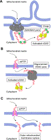
Mechanisms of mitochondrial DNA release. Mitochondrial DNA release is observed to occur via: (A) BAK and BAX pores which allow for mitochondrial inner membrane herniation through the outer membrane and escape of matrix components, including mtDNA, (B) mPTP channels in the inner mitochondrial membrane and oligomerized VDAC pores in the outer mitochondrial membrane, or (C) upon breakage of the outer mitochondrial membrane due to mPTP-mediated mitochondrial swelling. Once released, mitochondrial DNA can be sensed by DNA sensors. Cytosolic cGAS sensing is enhanced by TFAM.
Figure 4:
Figure 4:
Mechanisms of retrotransposon activation leading to cytosolic DNA generation. Upon transcriptional activation, polyadenylated (polyA) bicistronic LINE-1 (L1) mRNA encoding ORF1p and ORF2p is generated, exported to the cytoplasm, and translated. ORF1p and ORF2p bind to the mRNA forming a ribonucleoprotein (RNP) complex. Reverse transcription and L1 nuclear integration occurs via a mechanism known as target-primed reverse transcription (TPRT) that relies on annealing of poly(A) tail to thymidine nucleotides in nuclear DNA. Alternatively, reverse transcription potentially occurs in the cytoplasm. L1 cDNA accumulates in the cytoplasm in aging and disease, where it is sensed by nucleic acid sensors.
Figure 5:
Figure 5:
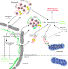
Cytosolic DNA species and sensing in aging and disease. Endogenous cytoDNA species are generated by distinct mechanisms but are sensed and processed by similar cellular pathways. Mechanisms assuring chromatin homeostasis (chromostasis) and DNA repair, as well as catabolism of cytoDNA, act to prevent excessive activation of innate immune signaling. Loss of these homeostatic processes is thought to contribute to inflammation and chronic diseases.

References

    1. Aarreberg LD, Esser-Nobis K, Driscoll C, Shuvarikov A, Roby JA, and Gale M Jr. (2019). Interleukin-1β Induces mtDNA Release to Activate Innate Immune Signaling via cGAS-STING. Mol Cell 74, 801–815.e806. 10.1016/j.molcel.2019.02.038. - DOI - PMC - PubMed
    1. Abe T, and Barber GN (2014). Cytosolic-DNA-mediated, STING-dependent proinflammatory gene induction necessitates canonical NF-kappaB activation through TBK1. J Virol 88, 5328–5341. 10.1128/JVI.00037-14. - DOI - PMC - PubMed
    1. Acosta JC, Banito A, Wuestefeld T, Georgilis A, Janich P, Morton JP, Athineos D, Kang TW, Lasitschka F, Andrulis M, et al. (2013). A complex secretory program orchestrated by the inflammasome controls paracrine senescence. Nat Cell Biol 15, 978–990. 10.1038/ncb2784. - DOI - PMC - PubMed
    1. Acosta JC, O’Loghlen A, Banito A, Guijarro MV, Augert A, Raguz S, Fumagalli M, Da Costa M, Brown C, Popov N, et al. (2008). Chemokine signaling via the CXCR2 receptor reinforces senescence. Cell 133, 1006–1018. S0092–8674(08)00619–3 [pii] 10.1016/j.cell.2008.03.038. - DOI - PubMed
    1. Aguado J, Chaggar HK, Gomez-Inclan C, Shaker MR, Leeson HC, Mackay-Sim A, and Wolvetang EJ (2021). Inhibition of the cGAS-STING pathway ameliorates the premature senescence hallmarks of Ataxia-Telangiectasia brain organoids. Aging Cell, e13468. 10.1111/acel.13468. - DOI - PMC - PubMed

Publication types