. 2022 Jan:109:125-134.
doi: 10.1016/j.neurobiolaging.2021.09.019. Epub 2021 Sep 30.

Abnormal tau in amyloid PET negative individuals

Affiliations

Abnormal tau in amyloid PET negative individuals

Bora Yoon et al. Neurobiol Aging. 2022 Jan.

Abstract

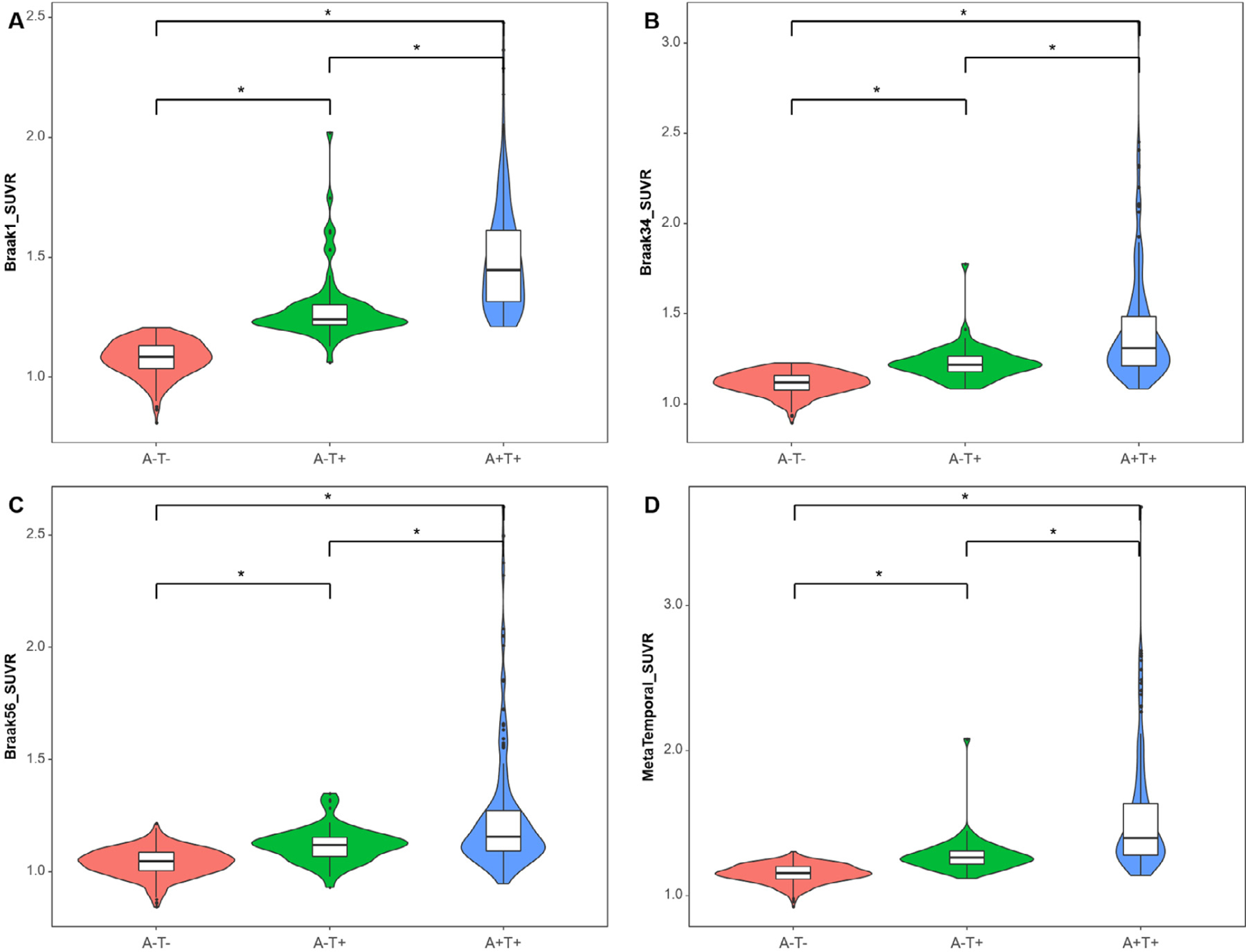
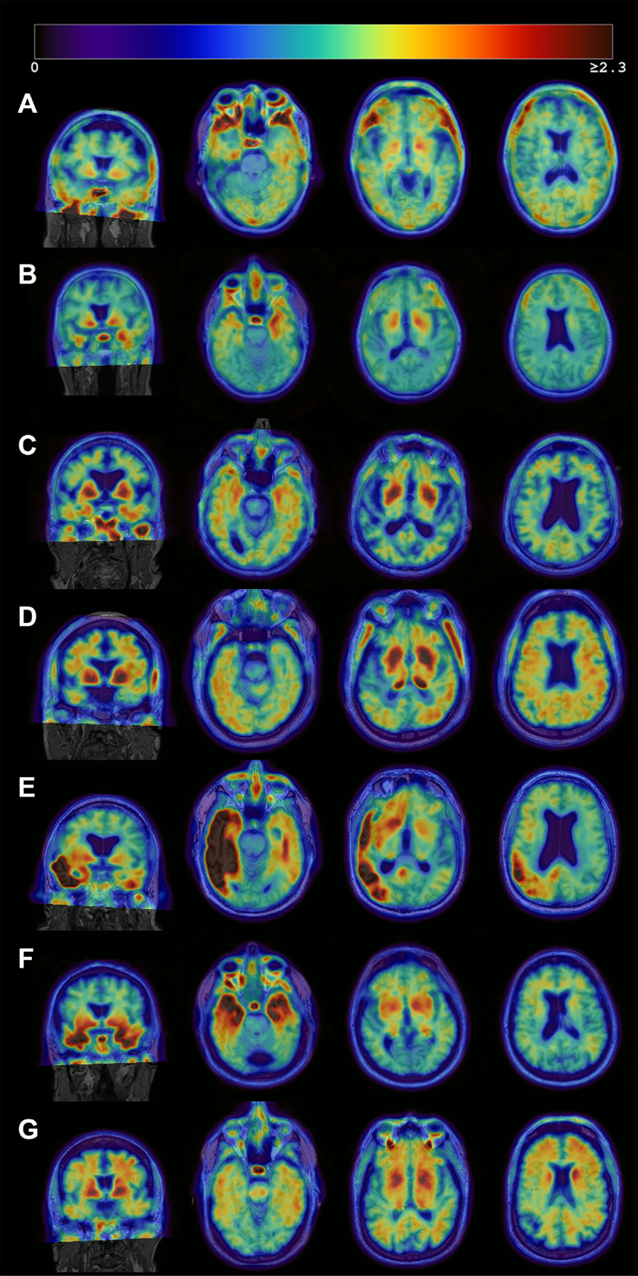
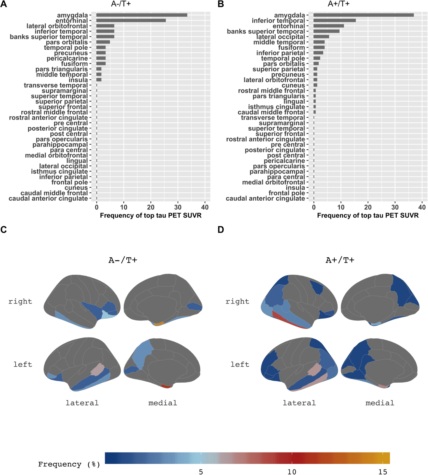
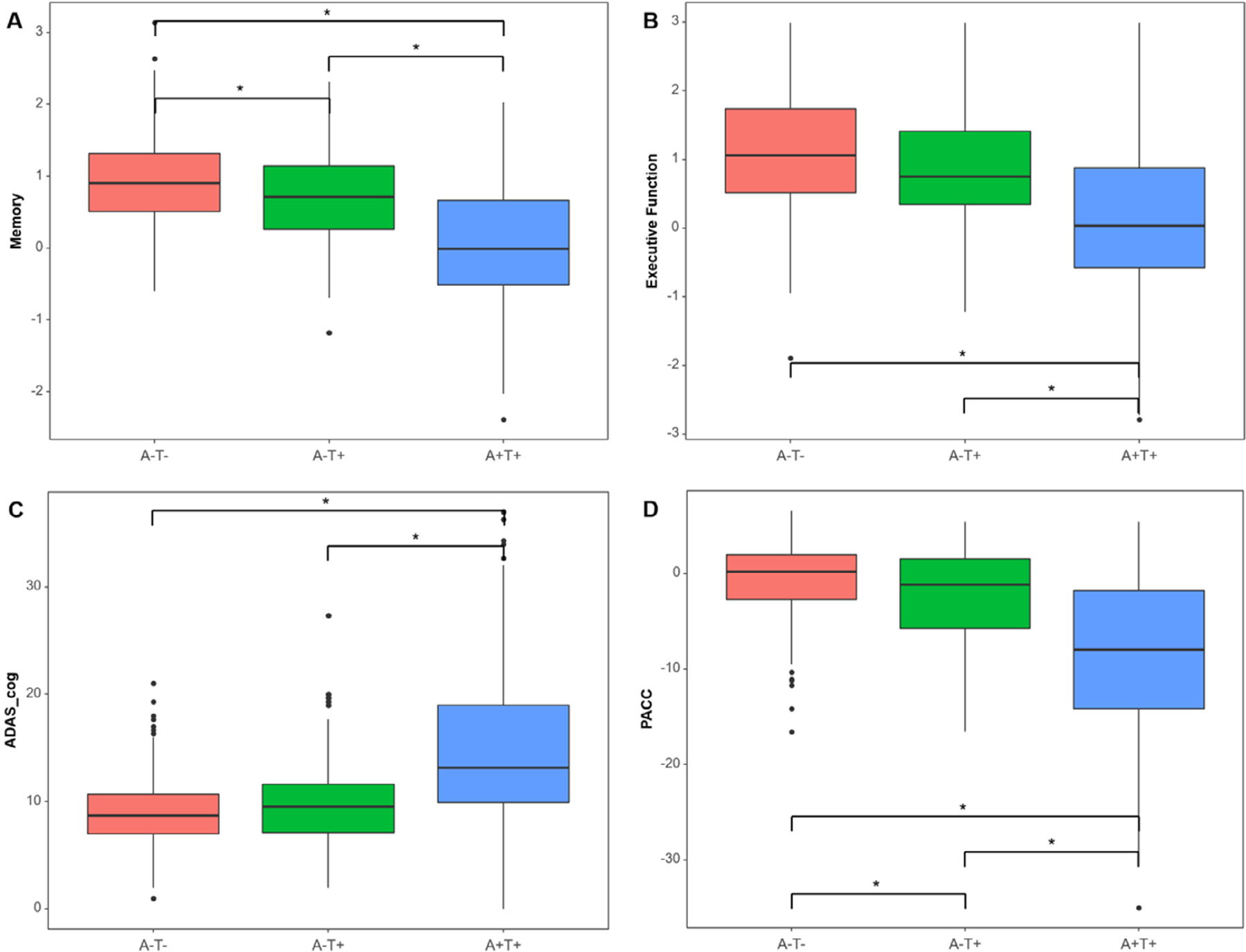
We examined the characteristics of individuals with biomarker evidence of tauopathy but without β-amyloid (Aβ) (A-T+) in relation to individuals with (A+T+) and without (A-T-) evidence of Alzheimer's disease (AD). We included 561 participants with Aβ and tau PET from the Alzheimer's Disease Neuroimaging Initiative (ADNI). We compared A-T- (n = 316), A-T+ (n = 63), and A+T+ (n = 182) individuals on demographics, amyloid, tau, hippocampal volumes, and cognition. A-T+ individuals were low on apolipoprotein E ɛ4 prevalence (17%) and had no evidence of subtly elevated brain Aβ within the negative range. The severity of tau deposition, hippocampal atrophy, and cognitive dysfunction in the A-T+ group was intermediate between A-T- and A+T+ (all p < 0.001). Tau uptake patterns in A-T+ individuals were heterogeneous, but approximately 29% showed tau deposition in the medial temporal lobe only, consistent with primary age-related tauopathy and an additional 32% showed a pattern consistent with AD. A-T+ individuals also share other features that are characteristic of AD such as cognitive impairment and neurodegeneration, but this group is heterogeneous and likely reflects more than one disorder.

Keywords: (18)F-flortaupir; Positron emission tomography; Primary age-related tauopathy; Tau; Tauopathies.


Conflict of interest statement

Disclosure statement

Dr. Yoon, Dr. Guo, Dr. Provost, Korman D, and Ward TJ report no disclosures relevant to the manuscript. Dr. Landau has served as a consultant to NeuroVison and Cortexyme. Dr. Jagust has served as a consultant to Genentech, Biogen, Novartis, CuraSen, Bioclinica, and Grifols.

Figures

Fig. 1.
Fig. 1.
Tau levels by Braak stage among 316 A-T- individuals, 63 A-T+, and 182 A+T+ individuals. Violin plots and Boxplots represent tau SUVR in grouped regions of interest corresponding to Braak 1 (A), Braak 34 (B), Braak 56 (C), and Metatemporal ROI (D) by AT group. The Y-axis is a tau SUVR value in each Braak stage. Regardless of Braak stages, the A-T+ group shows an intermediate level of FTP SUVR between A-T- and A+T+ groups. Asterisk () represents a statistically significant p-value (p < 0.001) in the analysis of covariance adjusted by age, sex, and diagnosis with Bonferroni correction. Abbreviations: FTP, 18F-flortaucipir; ROI, region of interests; SUVR, standard uptake value ratio.
Fig. 2.
Fig. 2.
Heterogeneous patterns of FTP binding in the A-T+ group. Each image consists of an MRI (grayscale) with the co-registered PET image overlaid. The images represent Negative or false-positive pattern (A), bilateral medial temporal binding pattern (B) or AD-like pattern (C, D), and atypical (E) or non-AD pattern (F, G). The right hemisphere is displayed on the right side. (A) No significant cortical binding on visual assessment. Likely false-positive quantification due to partial volume effect in the inferior temporal lobe, high off-target signal in the base of the skull, and non-specific signal in the white matter (68/female, MCI, MMSE 28). (B) Mild bilateral medial temporal FTP binding which could reflect PART (61/male, dementia, MMSE 23), (C) Mild bilateral medial and lateral temporal tracer retention which could represent PART or early AD (86/male, MCI, MMSE 28). (D) Mild lateral temporal, parietal, and frontal tracer retention in a pattern suggestive of AD (75/male, Normal, MMSE 26), (E) Intense asymmetric left-dominant parieto-temporal binding, highly suggestive of lvPPA due to AD despite amyloid negativity (71/male, Normal, MMSE 29), and (F) Intense binding in bilateral temporal poles, suggestive of MAPT mutation (68/male, MCI, MMSE 23) and (G) Mild to moderate tracer binding predominantly in the frontal white matter, suggestive of non-AD pathology (possible primary tauopathy) (68/male, MCI, MMSE 29). Abbreviations: AD, Alzheimer’s disease; FTP, 18F-flortaucipir; lvPPA, logopenic variant primary progressive aphasia; MAPT, microtubule-associated protein tau MCI, mild cognitive impairment; MMSE, mini-mental state examination; PART, primary age-related tauopathy.
Fig. 3.
Fig. 3.
The topography of tau deposition. In the bar charts of the A-T+ group (A) and the A+T+ group (B), the X-axis is the percentage of frequency that each region appeared as the highest tau SUVR (averaged across hemispheres). Detailed asymmetry index in the regions of each group were presented in Suppl. Fig. 4. Amygdala is the highest area of tau deposition in both the A-T+ group and the A+T+ group. The brain maps (C, D) demonstrate the distribution of tau deposition according to the frequency coded in the color bar below. Compared to the A-T+ group (C), the A+T+ group (D) shows a more widespread distribution of tau deposition extending to parietal, frontal, and occipital lobes, but similar patterns showing amygdala, temporal and orbitofrontal lobes. Abbreviations: SUVR, standard uptake value ratio.
Fig. 4.
Fig. 4.
Cognitive profile comparisons among 3 groups. Boxplots represent (A) memory composite score, (B) executive function composite score, (C) ADAS-cog, and (D) PACC by AT group. The Y-axis is a normalized value (z-scores) of each cognitive measure. The cognition of the A-T+ group is intermediate between the 3 groups. Asterisk () indicates a statistically significant p-value (p < 0.001) in the analysis of covariance adjusted by age, sex, and education with Bonferroni correction. Abbreviations: ADAS-cog, Alzheimer’s disease assessment scale-cognitive subscale; PACC, preclinical Alzheimer cognitive composite.

References

    1. Adams JN, Maass A, Harrison TM, Baker SL, Jagust WJ, 2019. Cortical tau deposition follows patterns of entorhinal functional connectivity in aging. Elife 8, e49132. - PMC - PubMed
    1. Altomare D, de Wilde A, Ossenkoppele R, Pelkmans W, Bouwman F, Groot C, van Maurik I, Zwan M, Yaqub M, Barkhof F, van Berckel BN, Teunissen CE, Frisoni GB, Scheltens P, van der Flier WM, 2019. Applying the ATN scheme in a memory clinic population. Neurology 93, e1635. - PubMed
    1. Bell WR, An Y, Kageyama Y, English C, Rudow GL, Pletnikova O, Thambisetty M, O’Brien R, Moghekar AR, Albert MS, Rabins PV, Resnick SM, Troncoso JC, 2019. Neuropathologic, genetic, and longitudinal cognitive profiles in primary age-related tauopathy (PART) and Alzheimer’s disease. Alzheimers Dement 15 (1), 8–16. - PMC - PubMed
    1. Besser LM, Crary JF, Mock C, Kukull WA, 2017. Comparison of symptomatic and asymptomatic persons with primary age-related tauopathy. Neurology 89 (16), 1707–1715. - PMC - PubMed
    1. Bilgel M, An Y, Helphrey J, Elkins W, Gomez G, Wong DF, Davatzikos C, Ferrucci L, Resnick SM, 2018. Effects of amyloid pathology and neurodegeneration on cognitive change in cognitively normal adults. Brain 141 (8), 2475–2485. - PMC - PubMed

Publication types

MeSH terms