. 2021 Nov 2;118(44):e2107500118.
doi: 10.1073/pnas.2107500118.

Mammalian sperm hyperactivation regulates navigation via physical boundaries and promotes pseudo-chemotaxis

Affiliations

Mammalian sperm hyperactivation regulates navigation via physical boundaries and promotes pseudo-chemotaxis

Meisam Zaferani et al. Proc Natl Acad Sci U S A. .

Abstract

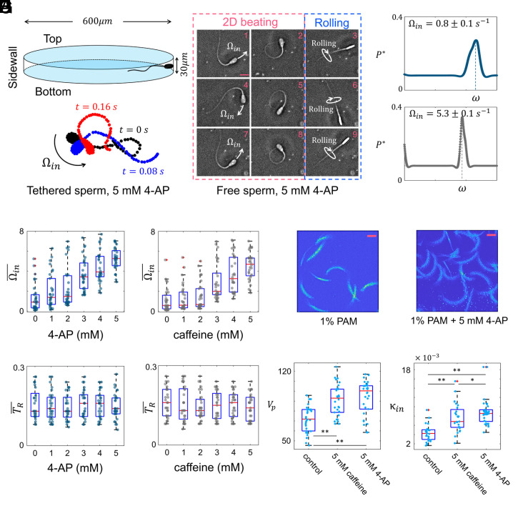
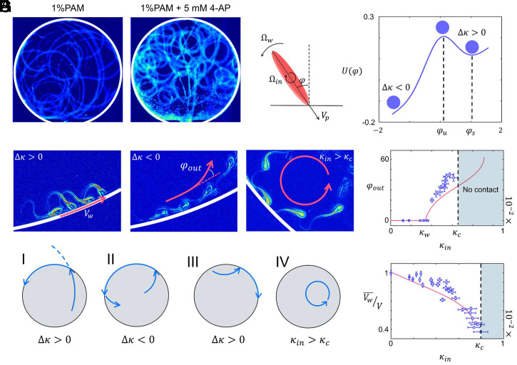
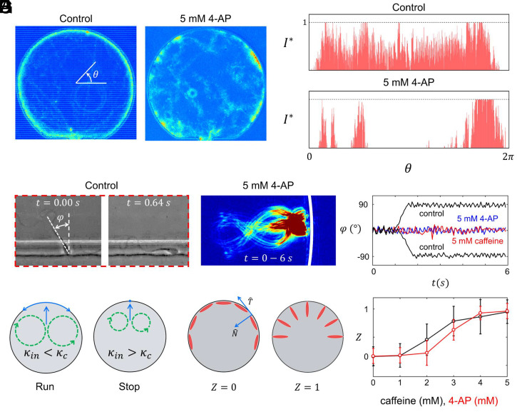
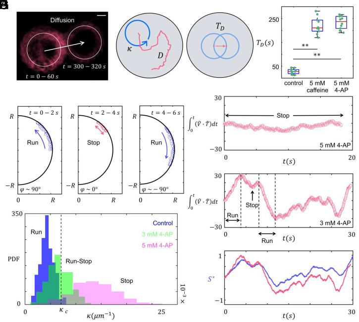
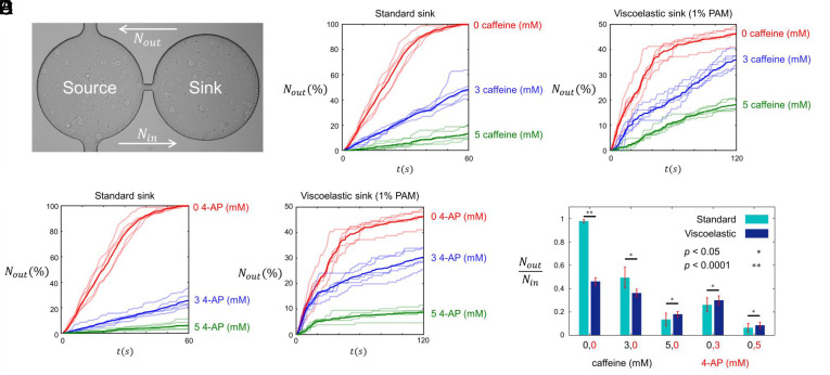
Mammalian sperm migration within the complex and dynamic environment of the female reproductive tract toward the fertilization site requires navigational mechanisms, through which sperm respond to the tract environment and maintain the appropriate swimming behavior. In the oviduct (fallopian tube), sperm undergo a process called "hyperactivation," which involves switching from a nearly symmetrical, low-amplitude, and flagellar beating pattern to an asymmetrical, high-amplitude beating pattern that is required for fertilization in vivo. Here, exploring bovine sperm motion in high-aspect ratio microfluidic reservoirs as well as theoretical and computational modeling, we demonstrate that sperm hyperactivation, in response to pharmacological agonists, modulates sperm-sidewall interactions and thus navigation via physical boundaries. Prior to hyperactivation, sperm remained swimming along the sidewalls of the reservoirs; however, once hyperactivation caused the intrinsic curvature of sperm to exceed a critical value, swimming along the sidewalls was reduced. We further studied the effect of noise in the intrinsic curvature near the critical value and found that these nonthermal fluctuations yielded an interesting "Run-Stop" motion on the sidewall. Finally, we observed that hyperactivation produced a "pseudo-chemotaxis" behavior, in that sperm stayed longer within microfluidic chambers containing higher concentrations of hyperactivation agonists.

Keywords: female reproductive tract; hyperactivation; mammalian sperm; navigation; sperm–wall interactions.


Conflict of interest statement

The authors declare no competing interest.

Figures

Fig. 1.
Fig. 1.
Characterization of hyperactivated motility. (A) The microfluidic reservoir. The diameter and height of reservoir are 600 and 30 µm, respectively. (B) Sequential images of free sperm flagellar beating in the standard solution with 5 mM 4-AP. The time between two consecutive images is 0.08 s. (C) Asymmetric flagellar beating of a tethered, 4-AP–treated sperm yields rotation about the tethering point. (D) Existence of a zeroth harmonic in the frequency domain results in the flagellar asymmetry and thus the sperm’s angular velocity about the tethering point. (E) Ωin¯ increases with the concentration of treatments. (F) TR¯ remained unchanged by 4-AP or caffeine. (G) Circular motion of sperm in viscoelastic (1% PAM) solution with and without 5 mM 4-AP. Rolling was suppressed in the viscoelastic solution. These images were obtained by combining consecutive images taken from sperm for 200 ms at 50 frames/s. (Scale bar, 50 µm.) Treatment with 5 mM caffeine or 4-AP increased sperm propulsive velocity (H) and circular path curvature in the viscoelastic solution (I). Units of Vp and κin are μms1 and μm1. *P value < 0.025 and **P value < 0.0001. These P values were obtained from two-tailed t tests, with adjustments for multiple comparisons (Bonferroni correction).
Fig. 2.
Fig. 2.
Effect of hyperactivation on sperm–sidewall interactions. (A) Time projection of sperm motion in viscoelastic solutions with and without 5 mM 4-AP. These images were obtained by combining frames acquired from sperm (25 frames/s) within circular reservoirs over 1-min periods. (B) Modeling sperm as a high–aspect ratio rod, the defining potential function of sperm–sidewall interactions is bistable, and sperm swimming behavior on the sidewall depends on the magnitude and sign of its intrinsic curvature (Ωin/Vp) at the contact point. x-axis, φ, Ωin, Vp, and Ωw  are depicted in the schematic. For Δκ > 0, a sperm swims along the sidewall (C), while for Δκ < 0, a sperm detaches after a temporary retention on the sidewall (D). (E) For κin>κc, no swimming along the sidewall occurs. Images shown in C, D, and E are obtained by combining four images taken from sperm at 100-ms intervals. (F) For Δκ<0, sperm velocity on the wall, and for Δκ > 0, detachment angle with respect to the intrinsic curvature. The solid red line is obtained from simulation, while blue dots represent experimental measurements. Error bars are used to indicate the estimated error in a measurement. The units for φout and κin are degrees and μm1. (G) Four categories of sperm–sidewall interactions.
Fig. 3.
Fig. 3.
Effect of hyperactivation on sperm–sidewall interactions. (A) Time projection of sperm motion in standard medium with and without 5 mM 4-AP. These images were obtained by combining frames acquired at (25 frames/sec) of sperm in circular reservoirs over 1-min periods. (B) The normalized optical density of the sperm layer formed around the circular sidewall with and without 5 mM 4-AP. The distribution of sperm around the sidewall was uniform in the control, whereas treatments with hyperactivation agonists yielded irregular accumulations along the sidewall. Similar results were obtained for 5 mM caffeine (shown in SI Appendix). (C) Rolling sperm reorientation after sidewall contact in the control medium. With 5 mM 4-AP (or caffeine), sperm maintained perpendicular orientation with respect to the sidewall. (D) Experimental measurements of sperm orientation with respect to the sidewall with and without treatment. (E) Rise of intrinsic curvature above the critical value yields Stop motion on the sidewall. Definition (F) and experimental measurements (G) of Z parameter at different concentrations of caffeine or 4-AP. Each point and error bar corresponds to three replicates.
Fig. 4.
Fig. 4.
Noise in the sperm intrinsic curvature. (A) Diffusivity of nonrolling sperm’s circular motion caused by the noise in the intrinsic curvature. This image was obtained by combining frames taken from sperm circular motion at 50 frames/s. (Scale bar, 50 µm.) (B and C) Definition of κ, D and TD. (D)  TD increased significantly with hyperactivation induced by 5 mM caffeine or 4-AP. **P value < 0.0001. These P values were obtained from two-tailed t tests, with adjustments for multiple comparisons (Bonferroni correction). (E) Sperm Run–Stop motion on the sidewall in a standard solution after treatment with 3 mM 4-AP, which incorporated change in the direction of swimming on the sidewall. Experimental measurements of sperm–sidewall orientation in Stop (5 mM 4-AP) (F) and Run–Stop (3 mM 4-AP) (G) types of motion. (H) Normalized sperm–sidewall orientation was proportional to the normalized arc length swum by sperm on the sidewall. (I) Probability density function of sperm intrinsic curvature with and without treatments. Noise promoted Run–Stop motion near the critical point.
Fig. 5.
Fig. 5.
Sperm pseudo-chemotaxis. (A) Two circular reservoirs (diameter: 600 µm) connected by a narrow channel (width: 40 µm). The left reservoir (Source) is filled with standard solution and no agonist; the right (Sink) contains treatments. Viscoelastic sinks contained 1% PAM. Nout as the cumulative percentage of sperm that exited the standard (B) and viscoelastic (C) sinks at 0, 3, and 5 mM caffeine. Nout as the cumulative percentage of sperm that exited the standard (D) and viscoelastic (E) sinks at 0, 3, and 5 mM 4-AP. All transparent lines indicate replicates, while the lines with solid colors indicate the means. (F) NoutNin ratios, corresponding to viscoelastic and standard sinks with different concentrations of caffeine or 4-AP. The P values were obtained from two-tailed t tests.

Similar articles

Cited by

References

    1. Eisenbach M., Giojalas L. C., Sperm guidance in mammals—An unpaved road to the egg. Nat. Rev. Mol. Cell Biol. 7, 276–285 (2006). - PubMed
    1. Suarez S. S., Pacey A. A., Sperm transport in the female reproductive tract. Hum. Reprod. Update 12, 23–37 (2006). - PubMed
    1. Miki K., Clapham D. E., Rheotaxis guides mammalian sperm. Curr. Biol. 23, 443–452 (2013). - PMC - PubMed
    1. Tung C. K., et al. ., Microgrooves and fluid flows provide preferential passageways for sperm over pathogen Tritrichomonas foetus. Proc. Natl. Acad. Sci. U.S.A. 112, 5431–5436 (2015). - PMC - PubMed
    1. Kantsler V., Dunkel J., Blayney M., Goldstein R. E., Rheotaxis facilitates upstream navigation of mammalian sperm cells. eLife 3, e02403 (2014). - PMC - PubMed

Publication types

LinkOut - more resources