. 2021 Nov 2;118(44):e2111031118.
doi: 10.1073/pnas.2111031118.

Kinetic proofreading of lipochitooligosaccharides determines signal activation of symbiotic plant receptors

Affiliations

Kinetic proofreading of lipochitooligosaccharides determines signal activation of symbiotic plant receptors

Kira Gysel et al. Proc Natl Acad Sci U S A. .

Abstract

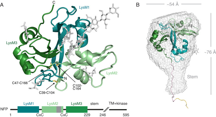
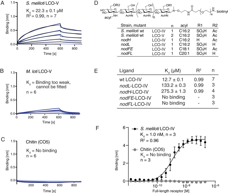
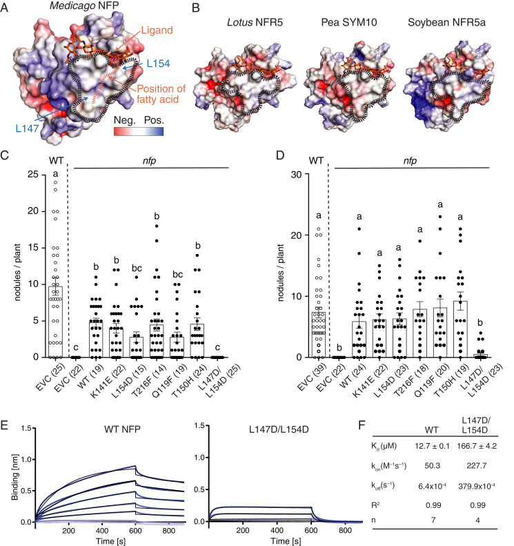
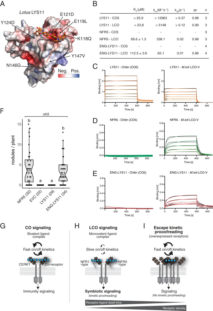
Plants and animals use cell surface receptors to sense and interpret environmental signals. In legume symbiosis with nitrogen-fixing bacteria, the specific recognition of bacterial lipochitooligosaccharide (LCO) signals by single-pass transmembrane receptor kinases determines compatibility. Here, we determine the structural basis for LCO perception from the crystal structures of two lysin motif receptor ectodomains and identify a hydrophobic patch in the binding site essential for LCO recognition and symbiotic function. We show that the receptor monitors the composition of the amphiphilic LCO molecules and uses kinetic proofreading to control receptor activation and signaling specificity. We demonstrate engineering of the LCO binding site to fine-tune ligand selectivity and correct binding kinetics required for activation of symbiotic signaling in plants. Finally, the hydrophobic patch is found to be a conserved structural signature in this class of LCO receptors across legumes that can be used for in silico predictions. Our results provide insights into the mechanism of cell-surface receptor activation by kinetic proofreading of ligands and highlight the potential in receptor engineering to capture benefits in plant-microbe interactions.

Keywords: LysM receptors; kinetic proofreading; legume symbiosis; lipochitooligosaccharide signaling; receptor–ligand interaction.


Conflict of interest statement

Competing interest statement: K.G., Z.B., L.H.M., S.R., J.S., and K.R.A., are inventors on patent application 62/718,186 submitted by Aarhus University that covers LysM receptors: genetically altered LysM receptors with altered agonist specificity and affinity. M.L., D.L., S.R., J.S., and K.R.A., are inventors on patent application 63/027,151 submitted by Aarhus University that covers LysM receptor motifs.

Figures

Fig. 1.
Fig. 1.
Structure of the NFP receptor ectodomain. (A) Cartoon representation of the NFP crystal structure with the three LysM domains colored as indicated in the schematic. Glycosylations are shown in gray and disulfide bridges in yellow. On the schematic representation of the protein, the position of the identified hydrophobic patch in LysM2 is indicated in dark gray. (B) Mesh representation of the NFP ab initio SAXS envelope with a rigid body fit of the ectodomain structure. The solution structure reveals a stem-like structure which is not visible in the crystal and a modeled possible configuration of the stem (light pink) and the hexahistidine tag (yellow). The overall dimensions are shown in angstrom (Å).
Fig. 2.
Fig. 2.
NFP has ligand specificity and directly monitors LCO decorations in BLI experiments. (A) NFP binding to S. meliloti LCO-V. (B) NFP binding to M. loti LCO-V is too weak and cannot be fitted. (C) NFP does not bind chitopentaose (CO5) in BLI experiments. A concentration range of analyte (100 to 1.56 µM) was used for each experiment. Experimental binding curves are represented in blue and fitting curves in black. The goodness of fit is described by the global fit R square of the mean value for each point. Numbers of replicates performed using independent protein preparations (n) are indicated. (D) Structure of biotinylated S. meliloti LCO-IV conjugate and overview of S. meliloti mutants associated with variations in LCO structure. S. meliloti LCO-IV has a tetrameric N-acetylglucosamine backbone, is O-sulfated on the reducing end, O-acetylated on the nonreducing terminal residue, and monoN-acylated by a hexadecadienoyl (C16:2) group. (E) BLI data showing NFP binding to S. meliloti LCO-IV variants. (F) Steady-state BLI data of full-length receptor binding at t = 595 s of association to immobilized LCO-IV and CO5, respectively. The binding follows a sigmoidal dose–response model with Kd = 1.0 ± 0.37 nM. Error bars indicate SD. A total of 16 twofold dilution series of analyte (200 to 0.0061 nM) were used for each experiment.
Fig. 3.
Fig. 3.
A hydrophobic patch in LysM2 is important for LCO binding and symbiotic signaling. (A) Molecular docking of a chitotetraose molecule (orange sticks) onto the structure of NFP. The surface of NFP is colored according to its electrostatic potential (±5 kT/e), and the hydrophobic patch is highlighted (black dashes). A possible position of the fatty acid chain on the hydrophobic patch is indicated (orange dashes). (B) Homology models of characterized LCO receptor ectodomains: L. japonicus NFR5, Pisum sativum (Pea) SYM10, and Glycine max (Soybean) NFR5α. All have a characteristic hydrophobic patch in LysM2. (C and D) Complementation analysis of NFP variants in an nfp Medicago background underlines that the hydrophobic patch is a prerequisite for functional symbiotic signaling. Columns represent mean nodule numbers after 49 d post infection (S. meliloti) (C) or 28 dpi (Sinorhizobium medicae) (D). Circles indicate individual counts. Empty circles: Medicago Jemalong wild-type background. Filled circles: nfp mutant background. EVC: empty vector control, WT: wild-type NFP. Error bars represent the SEM. Letters indicate statistical significance (ANOVA, Tukey, P < 0.05). Number of plants are indicated in parentheses. (E) BLI experiments of NFP WT and hydrophobic patch mutant (L147D/L154D) binding to S. meliloti LCO-IV. A concentration range of analyte (100 to 1.56 µM) was used for each experiment. Experimental binding curves are represented in blue and fitting curves in black. (F) Table summarizing the kinetic parameters for data in E. The goodness of fit is described by the global fit R square on the mean value of each point. Numbers of replicates performed using independent protein preparations (n) are indicated.
Fig. 4.
Fig. 4.
Engineering ligand specificity and binding kinetics enables LYS11 to support symbiotic signaling. (A) Electrostatic surface potential (±5 kT/e) representation of the crystal structure of LYS11. The chitin ligand of CERK1 is docked into the LysM2 chitin-binding groove. The hydrophobic patch and the proposed acyl chain binding site are indicated. Residues substituted in the engineered LYS11 version are indicated. (B) Table overview of ligand-binding parameters measured by BLI. A concentration range of analyte (100 to 1.56 µM) was used for each experiment. Experimental binding curves are represented in colors and fitting curves in black. The binding events were faster than the instrument sensitivity, so the values for LYS11 are given here as a minimal (kinetic parameters) or maximal (dissociation constant). (C) BLI experiments of LYS11 binding to CO5 and M. loti LCO-V. (D) BLI experiments of NFR5 binding to CO5 and M. loti LCO-V. (E) BLI experiments of engineered LYS11(ENG-LYS11) binding to CO5 and M. loti LCO-V. (F) Complementation analysis in an nfr5 Lotus background shows that the ligand binding site of LYS11 can be engineered to support symbiotic function. Columns represent mean nodule numbers after 35 dpi with M. loti. Circles indicate individual counts. EVC: empty vector control. Error bars represent the SEM. Letters indicate statistical significance (ANOVA, Tukey, P < 0.05). Numbers of plants are indicated in parentheses. (GI) Models of ligand perception and signaling in immunity and symbiosis. (G) Perception of a bivalent chitin ligand with fast kinetics by Arabidopsis CERK1/coreceptor complex leading to immunity signaling. (H) LCO perception by legume NFR1/NFR5 class receptors on a monovalent LCO ligand with slow binding and kinetic proofreading leading to symbiotic signaling. (I) Overexpression of LYS11 leads to a bypass of the kinetic proofreading mechanism despite fast on/off binding kinetics (40). Altering the hydrophobic patch in NFP leads to a faster binding kinetic and no symbiotic signaling (equal to receptor resting state) and engineering LYS11 with slow and specific LCO binding restores kinetics proofreading and symbiotic signaling even at low-receptor density when expressed from Nfr5 promoter.

References

    1. D’Haeze W., Holsters M., Nod factor structures, responses, and perception during initiation of nodule development. Glycobiology 12, 79R–105R (2002). - PubMed
    1. Cope K. R., et al. ., The ectomycorrhizal fungus Laccaria bicolor produces lipochitooligosaccharides and uses the common symbiosis pathway to colonize Populus roots. Plant Cell 31, 2386–2410 (2019). - PMC - PubMed
    1. Rush T. A., et al. ., Lipo-chitooligosaccharides as regulatory signals of fungal growth and development. Nat. Commun. 11, 3897 (2020). - PMC - PubMed
    1. Pueppke S. G., Broughton W. J., Rhizobium sp. strain NGR234 and R. fredii USDA257 share exceptionally broad, nested host ranges. Mol. Plant Microbe Interact. 12, 293–318 (1999). - PubMed
    1. Kelly S., Radutoiu S., Stougaard J., Legume LysM receptors mediate symbiotic and pathogenic signalling. Curr. Opin. Plant Biol. 39, 152–158 (2017). - PubMed

Publication types

LinkOut - more resources