Review
. 2021 Dec;78(23):7435-7449.
doi: 10.1007/s00018-021-03988-3. Epub 2021 Oct 30.

The lysosome as an imperative regulator of autophagy and cell death

Affiliations
Review

The lysosome as an imperative regulator of autophagy and cell death

Kewal Kumar Mahapatra et al. Cell Mol Life Sci. 2021 Dec.

Abstract

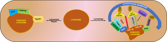
Lysosomes are single membrane-bound organelles containing acid hydrolases responsible for the degradation of cellular cargo and maintenance of cellular homeostasis. Lysosomes could originate from pre-existing endolysosomes or autolysosomes, acting as a critical juncture between autophagy and endocytosis. Stress that triggers lysosomal membrane permeabilization can be altered by ESCRT complexes; however, irreparable damage to the membrane results in the induction of a selective lysosomal degradation pathway, specifically lysophagy. Lysosomes play an indispensable role in different types of autophagy, including microautophagy, macroautophagy, and chaperone-mediated autophagy, and various cell death pathways such as lysosomal cell death, apoptotic cell death, and autophagic cell death. In this review, we discuss lysosomal reformation, maintenance, and degradation pathways following the involvement of the lysosome in autophagy and cell death, which are related to several pathophysiological conditions observed in humans.

Keywords: Autolysosome; Autophagic cell death; Autophagic lysosome reformation; Autophagy; Lysosome.


Conflict of interest statement

The authors declare that they have no conflict of interest with this manuscript.

Figures

Fig. 1
Fig. 1
Lysosomal reformation pathways. Lysosomal proteins are synthesized in the ER and transferred to the Golgi apparatus. Subsequently, they are transferred to the early endosome directly or through a constitutive secretory pathway via the plasma membrane. Following this, Rab5 is replaced by Rab7, which results in early endosome maturation for the formation of the late endosome. Late endosomes undergo fusion events, either homotypic (VAMP8) or heterotypic (VAMP7) with HOPS and SNARE complex to form hybrid endolysosomes. Then PIKfyve converts PI(3)P to PI(3,5)P2 to form the budded tubule from the endolysosome, which subsequently breaks to form protolysosome. Autophagy nucleation occurs with the source membrane derived from Golgi and ER. The phagophore is then elongated to form the autophagosome, which fuses with the lysosome to form the autolysosome. Subsequently, tubulation events occur with the help of the KIF5B and VPS34-UVRAG complex. PIP5K1B helps in converting PI(4)P to PI(4,5)P2. Then the budded tubule is fragmented with the help of DNM2 and PIP5K1A to form the protolysosome. The resultant protolysosomes undergo maturation to form the functional lysosomes, which are again available for the fusion events and degradation pathways
Fig. 2
Fig. 2
Lysosome repair or lysophagy. Stress response or excessive autophagy leads to the accumulation of exhausted lysosomes inside the cells. These lysosomal pools can be repaired via the recruitment of ALIX, CHMP4B, and ESCRT component Tsg101 in response to minor damage to the lysosomal membrane. In contrast, if the lysosomal membrane is extensively damaged, galactins sense this damage and are recruited onto the membrane-damaged site. Gal3 forms a complex with TRIM16 and recruits Ulk1 and ATG16L1 to the ubiquitination site, followed by the binding of p62/SQSTM1 to the LC3 positive phagophore membrane. Furthermore, K48 ubiquitination is regulated by the ubiquitin-directed AAA-ATPase VCP/p97 and the gene UBE2QL1. This complex binds to the autophagy adapter protein p62/SQSTM1 and subsequently binds to the LC3 positive phagophore membrane. Moreover, Gal8 can directly recruit NDP52 that can bind to LC3 positive phagophore to modulate lysophagy
Fig. 3
Fig. 3
Role of lysosome in autophagy. a Microautophagy: Cargo gets sequestered onto lysosome with the help of ESCRT and SNARE assembly, which helps in the incorporation of the cargo inside the lysosome. After internalization of the cargo, it gets degraded with the help of lysosomal hydrolases. b Macroautophagy: Autophagy is initiated following the inhibition of mTOR by response initiated to stress, which leads to activation of the ULK1-FIP200-ATG13 complex. Post activation of the initiation complex, Beclin-1 gets activated, which remains in the complex form with VPS15 and PI3KC3. Subsequently, the phagophore is elongated, followed by the formation of a matured autophagosome. The lysosome fuses with the autophagosome to form autolysosome, where the sequestered cargo from the autophagosome gets degraded, and nutrients are recycled back into the cell. c Chaperone-mediated autophagy: Hsc70 remains in a complex form with other co-chaperones bag1, hsp40, hip, hop, and hsp90. This complex helps in the identification of cargo containing a KFERQ sequence. Then, it binds to the LAMP2A monomer on the lysosome. In this instance, LAMP2A forms a multimeric structure leading to the formation of a translocon. Translocon provides a passage for the cargo to move inside the lysosome with the help of Lys-Hsc70. Subsequently, the cargo undergoes degradation
Fig. 4
Fig. 4
Role of lysosome in cell death: Lysosome remains intact with the help of proteins such as LAMP1, LAMP2, CD63, HSP70, and LIMP2. Any damage to these molecular players results in lysosomal membrane permeabilization. It leads to the release of cathepsins to the cytosol and activates the cell death signaling pathways. Cathepsin B, H, K, L, and S inhibit Bcl-2, Bcl-xL, Bak, Mcl-1, and BimEL; however, they cannot inhibit Bax and Bid. Activation of Bax and Bid leads to cell death. Furthermore, DRAM1 is regulated by p53 and modulates cell death by triggering mitochondrial outer membrane permeabilization, lysosomal membrane permeabilization, and autophagy activation. EVA1A activates ATG16L1 to induce autophagic cell death. An association of Beclin-1 with Bcl-2 results in the inhibition of autophagy, which leads to cell death. However, when BNIP3 sequesters Bcl-2, Beclin-1 is free to induce autophagy, thereby resulting in autophagic cell death. Moreover, ROS generation inside the cells helps in the peroxidation of lipids and proteins, leading to lysosomal membrane permeabilization and, in turn, cell death. In contrast, the ESCRT complex can repair the damaged lysosomal membrane and prevents cell death

References

    1. Koh J-Y, et al. Lysosomal dysfunction in proteinopathic neurodegenerative disorders: possible therapeutic roles of cAMP and zinc. Mol Brain. 2019;12(1):18–18. doi: 10.1186/s13041-019-0439-2. - DOI - PMC - PubMed
    1. Song Q, et al. The emerging roles of vacuolar-type ATPase-dependent Lysosomal acidification in neurodegenerative diseases. Transl Neurodegener. 2020;9(1):17–17. doi: 10.1186/s40035-020-00196-0. - DOI - PMC - PubMed
    1. Gyparaki M-T, Papavassiliou AG. Lysosome: the cell's ‘suicidal bag’ as a promising cancer target. Trends Mol Med. 2014;20(5):239–241. doi: 10.1016/j.molmed.2014.01.009. - DOI - PubMed
    1. Mahapatra KK, et al. Molecular interplay of autophagy and endocytosis in human health and diseases. Biol Rev. 2019;94(4):1576–1590. - PubMed
    1. Schulze H, Kolter T, Sandhoff K. Principles of lysosomal membrane degradation: Cellular topology and biochemistry of lysosomal lipid degradation. Biochim Biophys Acta (BBA) 2009;1793(4):674–683. doi: 10.1016/j.bbamcr.2008.09.020. - DOI - PubMed

LinkOut - more resources