Observational Study
. 2022 Feb;162(2):454-467.
doi: 10.1053/j.gastro.2021.10.029. Epub 2021 Oct 28.

Lower Serologic Response to COVID-19 mRNA Vaccine in Patients With Inflammatory Bowel Diseases Treated With Anti-TNFα

Affiliations
Observational Study

Lower Serologic Response to COVID-19 mRNA Vaccine in Patients With Inflammatory Bowel Diseases Treated With Anti-TNFα

Hadar Edelman-Klapper et al. Gastroenterology. 2022 Feb.

Abstract

Background & aim: Patients with inflammatory bowel diseases (IBD), specifically those treated with anti-tumor necrosis factor (TNF)α biologics, are at high risk for vaccine-preventable infections. Their ability to mount adequate vaccine responses is unclear. The aim of the study was to assess serologic responses to messenger RNA-Coronavirus Disease 2019 vaccine, and safety profile, in patients with IBD stratified according to therapy, compared with healthy controls (HCs).

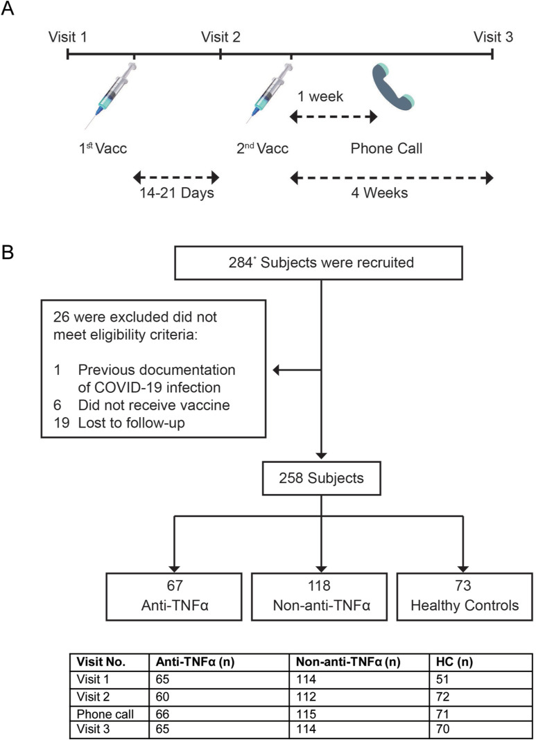
Methods: Prospective, controlled, multicenter Israeli study. Subjects enrolled received 2 BNT162b2 (Pfizer/BioNTech) doses. Anti-spike antibody levels and functional activity, anti-TNFα levels and adverse events (AEs) were detected longitudinally.

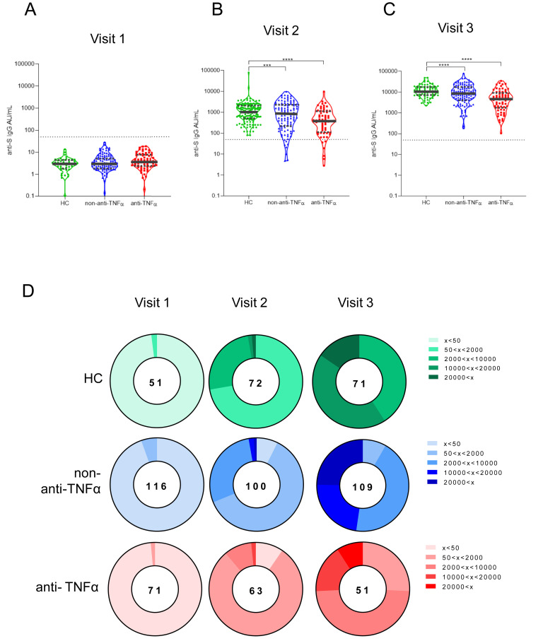
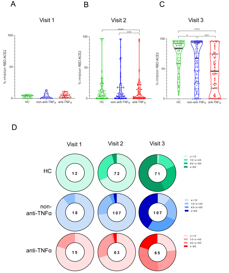
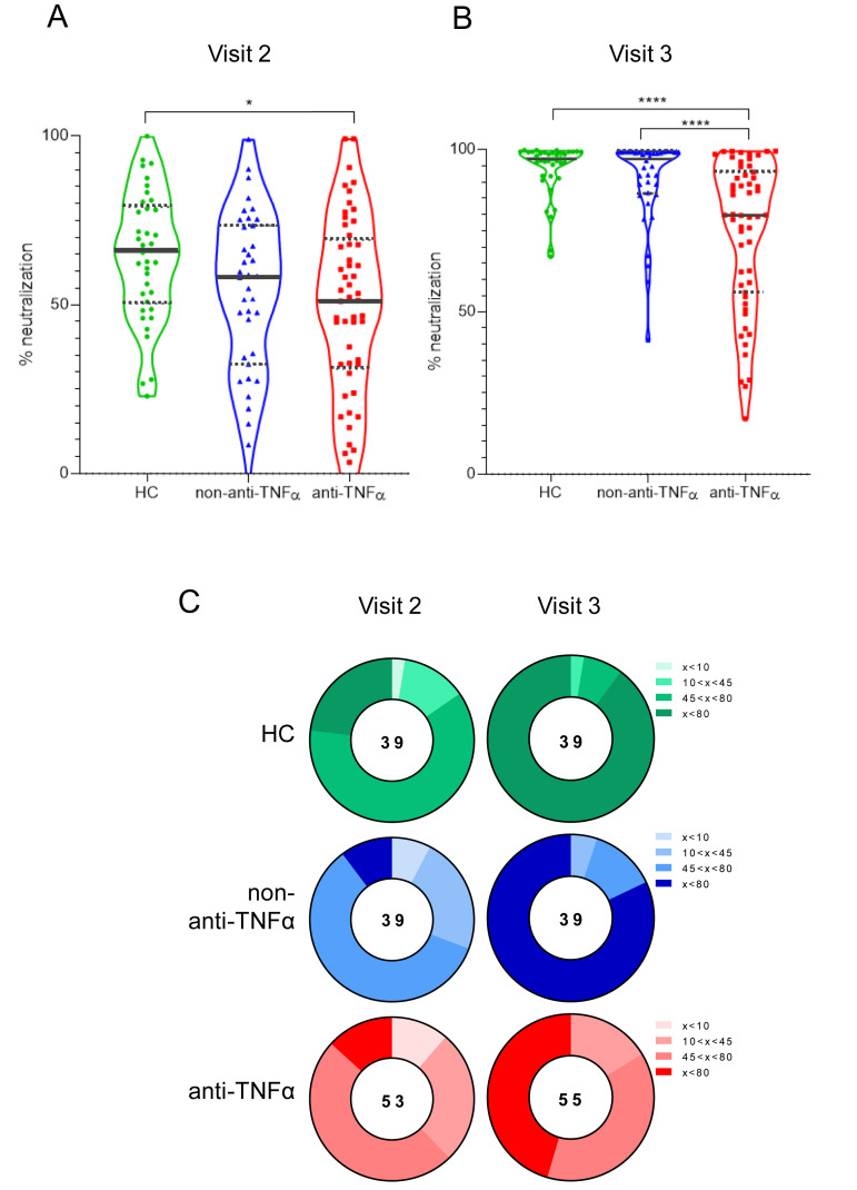
Results: Overall, 258 subjects: 185 IBD (67 treated with anti-TNFα, 118 non-anti-TNFα), and 73 HCs. After the first vaccine dose, all HCs were seropositive, whereas ∼7% of patients with IBD, regardless of treatment, remained seronegative. After the second dose, all subjects were seropositive, however anti-spike levels were significantly lower in anti-TNFα treated compared with non-anti-TNFα treated patients, and HCs (both P < .001). Neutralizing and inhibitory functions were both lower in anti-TNFα treated compared with non-anti-TNFα treated patients, and HCs (P < .03; P < .0001, respectively). Anti-TNFα drug levels and vaccine responses did not affect anti-spike levels. Infection rate (∼2%) and AEs were comparable in all groups. IBD activity was unaffected by BNT162b2.

Conclusions: In this prospective study in patients with IBD stratified according to treatment, all patients mounted serologic response to 2 doses of BNT162b2; however, its magnitude was significantly lower in patients treated with anti-TNFα, regardless of administration timing and drug levels. Vaccine was safe. As vaccine serologic response longevity in this group may be limited, vaccine booster dose should be considered.

Keywords: COVID-19; Serologic Response; Vaccine; mRNA-BNT162b2.


Figures

None
Graphical abstract
Figure 1
Figure 1
(A) Study protocol. Patients were enrolled at visit 1, before the first vaccine dose. Visit 2 was 14 to 21 days after the first but before the second vaccine dose. A week after the second vaccine dose, a phone call was made to evaluate AEs, and a visit 3 was 4 weeks after the second vaccine dose. In each visit, laboratory tests were performed, and questionnaires regarding disease severity and AEs were filled. (B) Patient disposition. The diagram represents all enrolled participants who were recruited before vaccination. ∗28 subjects were recruited at the second visit (after first vaccine dose but before the second one), mainly for logistic reasons. Most of them (22) were HCs. Number of subjects at each visit is detailed in the table below the diagram. Vacc, vaccine dose.
Figure 2
Figure 2
Patients with IBD treated with anti-TNFα have significantly reduced levels of anti-S antibodies. (A–C) Levels of anti-S antibodies in sera from HCs (shown in green), patients with IBD receiving non–anti-TNFα treatment (non–anti-TNFα, shown in blue), and patients with IBD receiving anti-TNFα treatment (anti-TNFα, shown in red). Antibodies were measured by the Abbott quantitative anti-S IgG kit. Visit 1 was before vaccination, visit 2 and visit 3, after first and second vaccine doses, respectively. Statistical analysis was carried out using independent-samples Kruskal-Wallis test. ∗∗∗P < .0005, ∗∗∗∗P < .0001. Black solid line denotes median, black dashed lines denote IQR 25–75. Dotted line represents the threshold for seroconversion (50 AU/mL). Specific GMCs and P values are in Supplementary Table 2. (D) Pie charts representing the fractions of patients at timepoint visits 1, 2, and 3, with anti-SARS-CoV-2 antibody levels as designated in the legend (A–C). Numbers in the middle of pies denote the total number of subjects tested in each group for every timepoint.
Figure 3
Figure 3
Patients with IBD treated with anti-TNFα have significantly reduced levels of anti-SARS-CoV-2 inhibiting antibodies. (A–C) Ability of serum from HCs (shown in green), patients with IBD receiving non–anti-TNFα treatment (non–anti-TNFα, shown in blue), and patients with IBD receiving anti-TNFα treatment (anti-TNFα, shown in red) to inhibit SARS-CoV-2 RBD binding to ACE2 receptor. Values measured by ELISA are presented as % inhibition (y axis), following vaccination. Visit 1 was before vaccination, and visit 2 and visit 3 were after first and second vaccine doses, respectively. Zero inhibition was set as the value of RBD without added sera. Statistical analysis was carried out using independent-samples Kruskal-Wallis test. ∗∗∗P < .0005, ∗∗∗∗P < .0001. At least 3 repetitions for every sample. Black solid line denotes median, black dashed lines denote IQR 25–75. Median percentage of inhibitions are in Supplementary Table 4. (D) Pie charts representing the fractions of patients at timepoint visits 1, 2, and 3, who developed none (<20%), low (20%<x<50%), medium (50%<x<80%), and high (>80%) SARS-CoV-2 RBD:ACE2 inhibition, based on (A–C). The numbers in the middle of the pies denote the total number of subjects tested in each group for every timepoint. Correlation between anti-S and inhibition responses are in Supplementary Figure 1.
Figure 4
Figure 4
Patients with IBD treated with anti-TNFα have significantly reduced levels of anti-SARS-CoV-2 neutralizing antibodies. (A, B) Sera, diluted to a final concentration of 1:200, were incubated with vesicular stomatitis virus-spike pseudo-particles (VSVΔGGFPSΔ19) for 1 hour at 37°C, before infecting ACE2 expressing human embryonic kidney 293 cells for 24 hours. The number of green fluorescent protein–positive cells was normalized and converted to a neutralization percentage in each sample, compared with the average of control samples. Visit 1 was before vaccination, visit 2 and visit 3 were after first and second vaccine doses, respectively. Statistical analysis was carried out using independent-samples Kruskal-Wallis test. ∗∗∗P < .0005, ∗∗∗∗P < .0001. Black solid line denotes median, black dashed lines denote IQR 25–75. (C) Pie charts representing the fractions of patients in timepoints visit 2 and visit 3, who developed none (<20%), low (20%<x<50%), medium (50%<x<80%), and high (>80%) SARS-CoV-2 RBD neutralizing antibodies, based on (A and B). Correlations between anti-S and neutralization, and between inhibition and neutralization, are in Supplementary Figures 2 and 3, respectively. HEK, human embryonic kidney; VSV; vesicular stomatitis virus.

References

    1. World Health Organization. Laboratory testing for 2019 novel coronavirus (2019-nCoV) in suspected human cases. Interim Guidance. World Health Organization; 2020.
    1. Polack F.P., Thomas S.J., Kitchin N., et al. Safety and efficacy of the BNT162b2 mRNA Covid-19 vaccine. N Engl J Med. 2020;383:2603–2615. - PMC - PubMed
    1. Jackson L.A., Anderson E.J., Rouphael N.G., et al. An mRNA vaccine against SARS-CoV-2 — preliminary report. N Engl J Med. 2020;383:1920–1931. - PMC - PubMed
    1. Rahier J.F., Magro F., Abreu C., et al. Second European evidence-based consensus on the prevention, diagnosis and management of opportunistic infections in inflammatory bowel disease. J Crohn’s Colitis. 2014;8:443–468. - PubMed
    1. Matsuoka K., Kobayashi T., Ueno F., et al. Evidence-based clinical practice guidelines for inflammatory bowel disease. J Gastroenterol. 2018;53:305–353. - PMC - PubMed

Publication types