. 2022 Feb;76(2):319-331.
doi: 10.1016/j.jhep.2021.10.009. Epub 2021 Oct 27.

Role of the IgG4-related cholangitis autoantigen annexin A11 in cholangiocyte protection

Affiliations

Role of the IgG4-related cholangitis autoantigen annexin A11 in cholangiocyte protection

Toni Herta et al. J Hepatol. 2022 Feb.

Abstract

Background & aims: Annexin A11 was identified as autoantigen in IgG4-related cholangitis (IRC), a B-cell driven disease. Annexin A11 modulates calcium-dependent exocytosis, a crucial mechanism for insertion of proteins into their target membranes. Human cholangiocytes form an apical 'biliary bicarbonate umbrella' regarded as defense against harmful hydrophobic bile acid influx. The bicarbonate secretory machinery comprises the chloride/bicarbonate exchanger AE2 and the chloride channel ANO1. We aimed to investigate the expression and function of annexin A11 in human cholangiocytes and a potential role of IgG1/IgG4-mediated autoreactivity against annexin A11 in the pathogenesis of IRC.

Methods: Expression of annexin A11 in human liver was studied by immunohistochemistry and immunofluorescence. In human control and ANXA11 knockdown H69 cholangiocytes, intracellular pH, AE2 and ANO1 surface expression, and bile acid influx were examined using ratio microspectrofluorometry, cell surface biotinylation, and 22,23-3H-glycochenodeoxycholic acid permeation, respectively. The localization of annexin A11-mEmerald and ANO1-mCherry was investigated by live-cell microscopy in H69 cholangiocytes after incubation with IRC patient serum containing anti-annexin A11 IgG1/IgG4-autoantibodies or disease control serum.

Results: Annexin A11 was strongly expressed in human cholangiocytes, but not hepatocytes. Knockdown of ANXA11 led to reduced plasma membrane expression of ANO1, but not AE2, alkalization of intracellular pH and uncontrolled bile acid influx. High intracellular calcium conditions led to annexin A11 membrane shift and colocalization with ANO1. Incubation with IRC patient serum inhibited annexin A11 membrane shift and reduced ANO1 surface expression.

Conclusion: Cholangiocellular annexin A11 mediates apical membrane abundance of the chloride channel ANO1, thereby supporting biliary bicarbonate secretion. Insertion is inhibited by IRC patient serum containing anti-annexin A11 IgG1/IgG4-autoantibodies. Anti-annexin A11 autoantibodies may contribute to the pathogenesis of IRC by weakening the 'biliary bicarbonate umbrella'.

Lay summary: We previously identified annexin A11 as a specific autoantigen in immunoglobulin G4-related cholangitis (IRC), a B-cell driven disease affecting the bile ducts. Human cholangiocytes are protected against harmful hydrophobic bile acid influx by a defense mechanism referred to as the 'biliary bicarbonate umbrella'. We found that annexin A11 is required for the formation of a robust bicarbonate umbrella. Binding of patient-derived annexin A11 autoantibodies inhibits annexin A11 function, possibly contributing to bile duct damage by weakening the biliary bicarbonate umbrella in patients with IRC.

Keywords: IgG4-related systemic disease; annexin A11; anoctamin-1; autoimmunity; bicarbonate umbrella; cholangiopathy; membrane trafficking.


Conflict of interest statement

Conflict of interest The authors declare no conflict of interest with regard to this work. Please refer to the accompanying ICMJE disclosure forms for further details.

Figures

Fig. 1.
Fig. 1.. Annexin A11 is expressed in the cytoplasm and the apical and basolateral plasma membrane of human cholangiocytes.
(A) Immunohistochemistry staining for annexin A11 in human liver tissue. (B) Cell surface biotinylation assay followed by immunoblotting for annexin A11 (observed molecular weight: 56 kDa) in sham and ANXA11 KD H69 cholangiocytes. Na+/K+ ATPase is used as loading control, GAPDH serves as proof for adequate separation of biotinylated plasma membrane proteins (no GAPDH) from intracellular proteins in the cell lysate. (C) Quantification of annexin A11 protein levels (7 cell samples of n = 3 independent experiments). (D) ANXA11 gene expression level in sham and ANXA11 KD H69 cholangiocytes (9 cell samples of n = 3 independent experiments). (E) Immunofluorescence staining for annexin A11 in human liver tissue. ▶ basolateral cholangiocyte cell membrane, # bile duct lumen. Data are presented as median with interquartile range. Levels of significance: ***p <0.001, ****p <0.0001 (Mann-Whitney U test (C), unpaired t test (D)). KD, knockdown; NB, non-biotin control of sham H69 cholangiocytes.
Fig. 2.
Fig. 2.. Plasma membrane expression of annexin A11 is pH-sensitive in H69 cholangiocytes.
(A) Cell surface biotinylation assay followed by immunoblotting for annexin A11 in sham H69 cholangiocytes after treatment with 20 mM sodium chloride (control) or sodium acetate (Acetate) for 10 minutes. Na+/K+ ATPase is used as loading control, GAPDH serves as proof for adequate separation of biotinylated plasma membrane proteins (no GAPDH) from total cell lysate. (B) Quantification of protein levels (12 cell samples of n = 6 independent experiments). (C) Intracellular pH tracing in sham H69 cholangiocytes without (w/o) and with (w/) 20 mM sodium acetate treatment. (D) Baseline intracellular pH of sham and ANXA11 KD H69 cholangiocytes (n = 4 independent experiments). (E) Representative intracellular pH tracing of sham and ANXA11 KD H69 cholangiocytes in HBSS and Cl-free HBSS. Data are presented as median with interquartile range (B, D) or average of duplicate (C, E). Levels of significance: *p <0.05, **p <0.01 (Mann-Whitney U test (B), paired t-test (D)). HBSS, Hanks’ balanced salt solution; KD, knockdown; NB, non-biotin control of sham H69 cholangiocytes.
Fig. 3.
Fig. 3.. Annexin A11 is required for ANO1, but not AE2 membrane localization in human cholangiocytes.
Cell surface biotinylation assay followed by immunoblotting for (A) ANO1 (observed molecular weight: 75 kDa) or (B) AE2 (observed molecular weight: 150 kDa) in sham and ANXA11 KD H69 cholangiocytes. Na+/K+ ATPase is used as loading control, GAPDH serves as proof for adequate separation of biotinylated plasma membrane proteins (no GAPDH) from total cell lysate. (C, D) Quantification of protein levels (6 cell samples of n = 3 independent experiments). (E) Baseline intracellular pH of sham and ANO1 KD H69 cholangiocytes (n = 3 independent experiments). (F) Representative full trace of baseline intracellular pH in sham and ANO1 KD H69 cholangiocytes. (G) Immunofluorescence staining for annexin A11, AE2 and ANO1 in human liver. (H) Pearson’s correlation coefficients (7 bile ducts in n = 3 livers). Data are presented as median with interquartile range. Levels of significance: **p <0.01, n.s. not significant (Mann-Whitney U test (C, D), paired t test (E)). KD, knockdown; NB, non-biotin control of sham H69 cholangiocytes.
Fig. 4.
Fig. 4.. Colocalization of annexin A11 and ANO1 is stimulated by elevated intracellular free Ca2+ levels in H69 cholangiocytes.
(A) Live-cell imaging of ionomycin treated (10 μM for 15 minutes) annexin A11-mEmerald-over-expressing and ANO1-mCherry-transfected H69 cholangiocytes. (B) Pearson’s correlation coefficient in the region of interest (defined as cells showing annexin A11-mEmerald localization shift and ANO1-mCherry expression) (6 regions of interest of n = 3 independent experiments). Data are presented as median with interquartile range. Level of significance: ****p <0.0001 (unpaired t test).
Fig. 5.
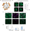
Fig. 5.. Serum of a patient with IRC containing anti-annexin A11 IgG1/IgG4-autoantibodies, but not of patients with PSC, inhibits the Ca2+-dependent membrane shift of annexin A11 and reduces plasma membrane localization of ANO1.
(A) IgG1/IgG4-autoantibody binding sites on annexin A11 (orange) and Ca2+-binding domains (cyan) as predicted using the ElliPro software tool. (B) Ionomycin-treated (50 μM for 15 minutes) annexin A11-mEmerald-overexpressing H69 cholangiocytes after 3 days of incubation with 20% PSC or IRC patient serum. Proportion of cells showing annexin A11-mEmerald localization shift (7 confocal pictures of n = 3 independent experiments). (C) ANO1-mCherry plasma membrane localization in H69 cholangiocytes after 6 days of incubation with 20% PSC or IRC patient serum. (D) Semiquantitative scoring of ANO1-mCherry plasma membrane localization (1 scorer, n = 3 independent experiments; see also Fig. S5 for scorer 2 and 3). Data are presented as median with interquartile range. Level of significance: **p <0.01, ****p <0.0001 (unpaired t test (B), two-way ANOVA (D)). IRC, IgG4-related cholangitis; PSC, primary sclerosing cholangitis.
Fig. 6.
Fig. 6.. Knockdown of ANXA11 and ANO1 increases plasma membrane permeability for glycine-conjugated bile acids in H69 cholangiocytes.
22,23-3H-GCDC permeation assay in sham and (A) ANXA11 KD or (C) ANO1 KD H69 cholangiocytes. (B, D) Quantification of 22,23-3H-GCDC permeation (9 cell samples of n = 3 independent experiments (B) or 12 cell samples of n = 4 independent experiments (D)). Data are presented as median with interquartile range. Level of significance: *p <0.05, **p <0.01, ****p <0.0001, n.s. not significant (unpaired t test). DPM, disintegrations per minute; GCDC, glycine-conjugated chenodeoxycholic acid; KD, knockdown.

References

    1. Stone JH, Zen Y, Deshpande V. IgG4-related disease. N Engl J Med 2012;366:539–551. - PubMed
    1. Ghazale A, Chari ST, Zhang L, Smyrk TC, Takahashi N, Levy MJ, et al. Immunoglobulin G4-associated cholangitis: clinical profile and response to therapy. Gastroenterology 2008;134:706–715. - PubMed
    1. Maillette de Buy Wenniger LJ, Doorenspleet ME, Klarenbeek PL, Verheij J, Baas F, Elferink RP, et al. Immunoglobulin G4+ clones identified by next-generation sequencing dominate the B cell receptor repertoire in immunoglobulin G4 associated cholangitis. Hepatology 2013;57:2390–2398. - PubMed
    1. Doorenspleet ME, Hubers LM, Culver EL, Maillette de Buy Wenniger LJ, Klarenbeek PL, Chapman RW, et al. Immunoglobulin G4(+) B-cell receptor clones distinguish immunoglobulin G 4-related disease from primary sclerosing cholangitis and biliary/pancreatic malignancies. Hepatology 2016;64:501–507. - PMC - PubMed
    1. Mattoo H, Mahajan VS, Della-Torre E, Sekigami Y, Carruthers M, Wallace ZS, et al. De novo oligoclonal expansions of circulating plasma-blasts in active and relapsing IgG4-related disease. J Allergy Clin Immunol 2014;134:679–687. - PMC - PubMed

Publication types