. 2021 Dec;12(2):9561-9574.
doi: 10.1080/21655979.2021.1997561.

Protein tyrosine phosphatase receptor type O (PTPRO) knockdown enhances the proliferative, invasive and angiogenic activities of trophoblast cells by suppressing ER resident protein 44 (ERp44) expression in preeclampsia

Affiliations

Protein tyrosine phosphatase receptor type O (PTPRO) knockdown enhances the proliferative, invasive and angiogenic activities of trophoblast cells by suppressing ER resident protein 44 (ERp44) expression in preeclampsia

Yang Yang et al. Bioengineered. 2021 Dec.

Abstract

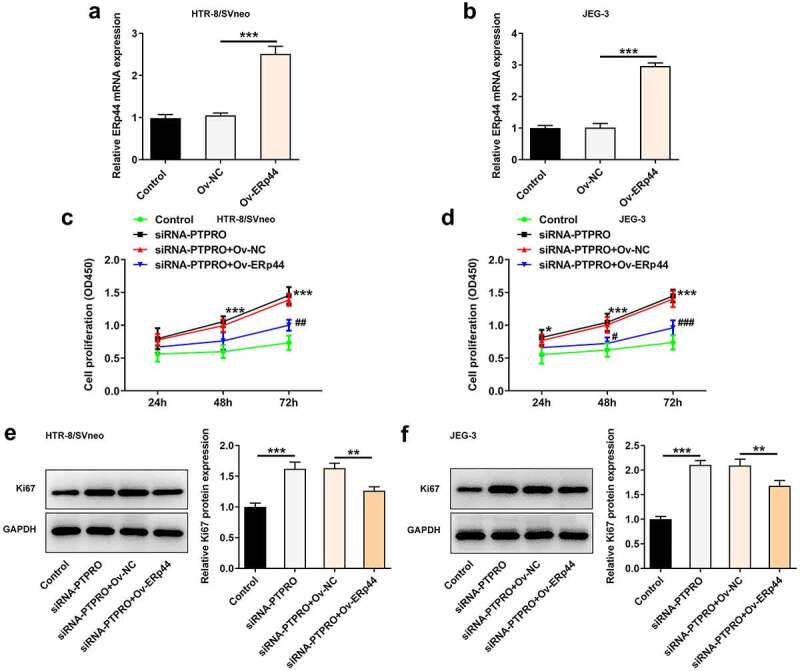
Preeclampsia (PE), a pregnancy-specific syndrome, is the primary cause of maternal mortality. This work was designed to investigate the specific functions of PTPRO/ ERp44 in the biological behaviors of trophoblast cells and elucidate the underlying molecular mechanism. Constructed siRNA-PTPRO and ERp44 overexpression plasmids were transfected into HTR-8/SVneo and JEG-3 cells for further functional experiments. Subsequently, the proliferation and invasion of trophoblast cells were identified by performing CCK-8, flow cytometry and transwell assay. In addition, tube formation assay was employed to estimate the angiogenic ability of HUVECs incubated with the conditioned media (CM) of HTR-8/SVneo or JEG-3 cells. Importantly, the interaction between PTPRO and ERp44 was analyzed through Co-IP. In the current investigation, it was discovered that downregulation of PTPRO notably facilitated the proliferation and invasion of trophoblast cells and induced a stronger in vitro angiogenesis. Moreover, PTPRO interacted with ERp44 to regulate ERp44 expression. ERp44 overexpression suppressed the proliferative, invasive and angiogenic activities of trophoblast cells. As a result, functions of PTPRO knockdown in the biological behaviors of trophoblast cells were partially abrogated upon elevation of ERp44. To sum up, this current research systematically evidenced that PTPRO could regulate the biological behaviors of trophoblast cells by modulating ERp44. Findings may contribute to a novel therapeutic strategy for PE.

Keywords: ERp44; PTPRO; preeclampsia; trophoblast cells.


Conflict of interest statement

No potential conflict of interest was reported by the author(s).

Figures

Figure 1.
Figure 1.
Downregulation of PTPRO promoted the proliferation of trophoblast cells. HTR-8/SVneo and JEG-3 cells were transfected with siRNA-PTPRO-1/2 or siRNA-NC. (a) RT-qPCR was employed to detect the mRNA level of PTPRO in HTR-8/SVneo cells. (b) Western blot analysis was employed to detect the protein level of PTPRO in HTR-8/SVneo cells. (c) RT-qPCR was employed to detect the mRNA level of PTPRO in JEG-3 cells. (d) Western blot analysis was employed to detect the protein level of PTPRO in JEG-3 cells. (e) CCK-8 assay was employed to assess the proliferation of HTR-8/SVneo cells. (f) CCK-8 assay was employed to assess the proliferation of JEG-3 cells. (g) Western blot analysis was employed to detect the protein level of Ki67 in HTR-8/SVneo cells. (h) Western blot analysis was employed to detect the protein level of Ki67 in JEG-3 cells. **p < 0.01, ***p < 0.001
Figure 2.
Figure 2.
Downregulation of PTPRO promoted cell cycle progression. HTR-8/SVneo and JEG-3 cells were transfected with siRNA-PTPRO or siRNA-NC. (a, b) Flow cytometry was adopted to evaluate the cell cycle distribution of HTR-8/SVneo cells. (c, d) Flow cytometry was adopted to evaluate the cell cycle distribution of JEG-3 cells. ***p < 0.001
Figure 3.
Figure 3.
Downregulation of PTPRO facilitated the invasion of trophoblast cells. HTR-8/SVneo and JEG-3 cells were transfected with siRNA-PTPRO or siRNA-NC. (a, b) Transwell assay was performed to evaluate the invasion of HTR-8/SVneo cells. (c, d) Transwell assay was performed to evaluate the invasion of JEG-3 cells. (e) RT-qPCR was employed to detect the mRNA levels of MMP2 and MMP9 in HTR-8/SVneo cells. (f) Western blot analysis was employed to detect the protein levels of MMP2 and MMP9 in HTR-8/SVneo cells. (g) RT-qPCR was employed to detect the mRNA levels of MMP2 and MMP9 in JEG-3 cells. (h) Western blot analysis was employed to detect the protein levels of MMP2 and MMP9 in JEG-3 cells. **p < 0.01, ***p < 0.001
Figure 4.
Figure 4.
Downregulation of PTPRO induced a stronger in vitro angiogenesis. HTR-8/SVneo and JEG-3 cells were transfected with siRNA-PTPRO or siRNA-NC. HUVECs were incubated with the conditioned media (CM) of HTR-8/SVneo or JEG-3 cells. (a-c) The angiogenic activity of HUVECs incubated with the conditioned media (CM) of HTR-8/SVneo cells was tested using an in vitro tube formation assay. (d-f) The angiogenic activity of HUVECs incubated with the conditioned media (CM) of JEG-3 cells was tested using an in vitro tube formation assay. (g) Western blot analysis was employed to detect the protein levels of VEGF, ET-1 and sFlt-1 in HUVECs incubated with the conditioned media (CM) of HTR-8/SVneo cells. (h) Western blot analysis was employed to detect the protein levels of VEGF, ET-1 and sFlt-1 in HUVECs incubated with the conditioned media (CM) of JEG-3 cells. **p < 0.01, ***p < 0.001
Figure 5.
Figure 5.
PTPRO interacted with ERp44 to participate in PE progression. (a-d) Co-IP assays were carried out to determine the interaction between PTPRO and ERp44. (e, f) HTR-8/SVneo and JEG-3 cells were transfected with siRNA-PTPRO or siRNA-NC. Western blot analysis was employed to detect the protein level of ERp44 in HTR-8/SVneo and JEG-3 cells. ***p < 0.001
Figure 6.
Figure 6.
Downregulation of PTPRO promoted the proliferation of trophoblast cells by suppressing ERp44 expression. (a, b) HTR-8/SVneo and JEG-3 cells were transfected with Ov-ERp44 or Ov-NC. RT-qPCR was employed to detect the mRNA level of ERp44 in HTR-8/SVneo and JEG-3 cells. (c, d) HTR-8/SVneo and JEG-3 cells were transfected with siRNA-PTPRO or co-transfected with siRNA-PTPRO and Ov-ERp44. CCK-8 assay was employed to assess the proliferation of HTR-8/SVneo and JEG-3 cells. (e, f) HTR-8/SVneo and JEG-3 cells were transfected with siRNA-PTPRO or co-transfected with siRNA-PTPRO and Ov-ERp44. Western blot analysis was employed to detect the protein level of Ki67 in HTR-8/SVneo and JEG-3 cells. *p < 0.05, **p < 0.01, ***p < 0.001; #p < 0.05, ##p < 0.01, ###p < 0.001
Figure 7.
Figure 7.
Downregulation of PTPRO promoted cell cycle progression by suppressing ERp44 expression. HTR-8/SVneo and JEG-3 cells were transfected with siRNA-PTPRO or co-transfected with siRNA-PTPRO and Ov-ERp44. (a, b) Flow cytometry was adopted to evaluate the cell cycle distribution of HTR-8/SVneo cells. (c, d) Flow cytometry was adopted to evaluate the cell cycle distribution of JEG-3 cells. *p < 0.05, **p < 0.01, ***p < 0.001
Figure 8.
Figure 8.
Downregulation of PTPRO facilitated the invasion of trophoblast cells by suppressing ERp44 expression. HTR-8/SVneo and JEG-3 cells were transfected with siRNA-PTPRO or co-transfected with siRNA-PTPRO and Ov-ERp44. (a, b) Transwell assay was performed to evaluate the invasion of HTR-8/SVneo cells. (c, d) Transwell assay was performed to evaluate the invasion of JEG-3 cells. (e) RT-qPCR was employed to detect the mRNA levels of MMP2 and MMP9 in HTR-8/SVneo cells. (f) Western blot analysis was employed to detect the protein levels of MMP2 and MMP9 in HTR-8/SVneo cells. (g) RT-qPCR was employed to detect the mRNA levels of MMP2 and MMP9 in JEG-3 cells. (h) Western blot analysis was employed to detect the protein levels of MMP2 and MMP9 in JEG-3 cells. * p < 0.05, **p < 0.01, ***p < 0.001
Figure 9.
Figure 9.
Downregulation of PTPRO induced a stronger in vitro angiogenesis by suppressing ERp44 expression. HTR-8/SVneo and JEG-3 cells were transfected with siRNA-PTPRO or co-transfected with siRNA-PTPRO and Ov-ERp44. HUVECs were incubated with the conditioned media (CM) of HTR-8/SVneo or JEG-3 cells. (a-c) The angiogenic activity of HUVECs incubated with the conditioned media (CM) of HTR-8/SVneo cells was tested using an in vitro tube formation assay. (d-f) The angiogenic activity of HUVECs incubated with the conditioned media (CM) of JEG-3 cells was tested using an in vitro tube formation assay. (g) Western blot analysis was employed to detect the protein levels of VEGF, ET-1 and sFlt-1 in HUVECs incubated with the conditioned media (CM) of HTR-8/SVneo cells. (h) Western blot analysis was employed to detect the protein levels of VEGF, ET-1 and sFlt-1 in HUVECs incubated with the conditioned media (CM) of JEG-3 cells. **p < 0.01, ***p < 0.001

References

    1. Zheng L, Song J, Tang R, et al. MicroRNA‑524‑5p regulates the proliferation and invasion of HTR-8/SVneo trophoblasts by targeting NUMB in the Notch signaling pathway. Mol Med Rep. 2021;23(6):6. - PMC - PubMed
    1. Zhou C, Zou QY, Li H, et al. Preeclampsia downregulates MicroRNAs in fetal endothelial cells: roles of miR-29a/c-3p in endothelial function. J Clin Endocrinol Metab. 2017;102(9):3470–3479. - PMC - PubMed
    1. Hu S, Li J, Tong M, et al. MicroRNA‑144‑3p may participate in the pathogenesis of preeclampsia by targeting Cox‑2. Mol Med Rep. 2019;19(6):4655–4662. - PMC - PubMed
    1. Travaglino A, Raffone A, Saccone G, et al. Placental morphology, apoptosis, angiogenesis and epithelial mechanisms in early-onset preeclampsia. Eur J Obstet Gynecol Reprod Biol. 2019;234:200–206. - PubMed
    1. Hou J, Xu J, Jiang R, et al. Estrogen-sensitive PTPRO expression represses hepatocellular carcinoma progression by control of STAT3. Hepatology. 2013;57(2):678–688. - PubMed

Publication types

MeSH terms

Substances