. 2021 Jan-Dec;13(1):1981202.
doi: 10.1080/19420862.2021.1981202.

Preclinical characterization of bemarituzumab, an anti-FGFR2b antibody for the treatment of cancer

Affiliations

Preclinical characterization of bemarituzumab, an anti-FGFR2b antibody for the treatment of cancer

Hong Xiang et al. MAbs. 2021 Jan-Dec.

Abstract

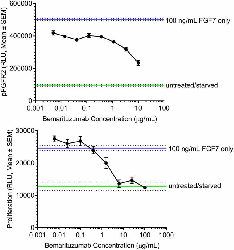
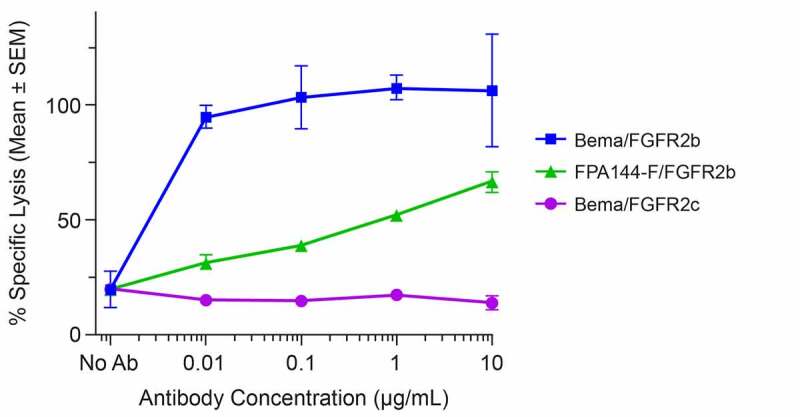
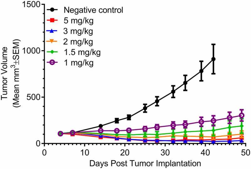
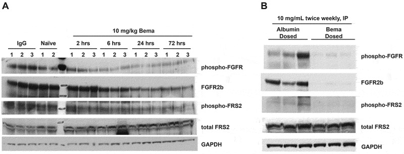
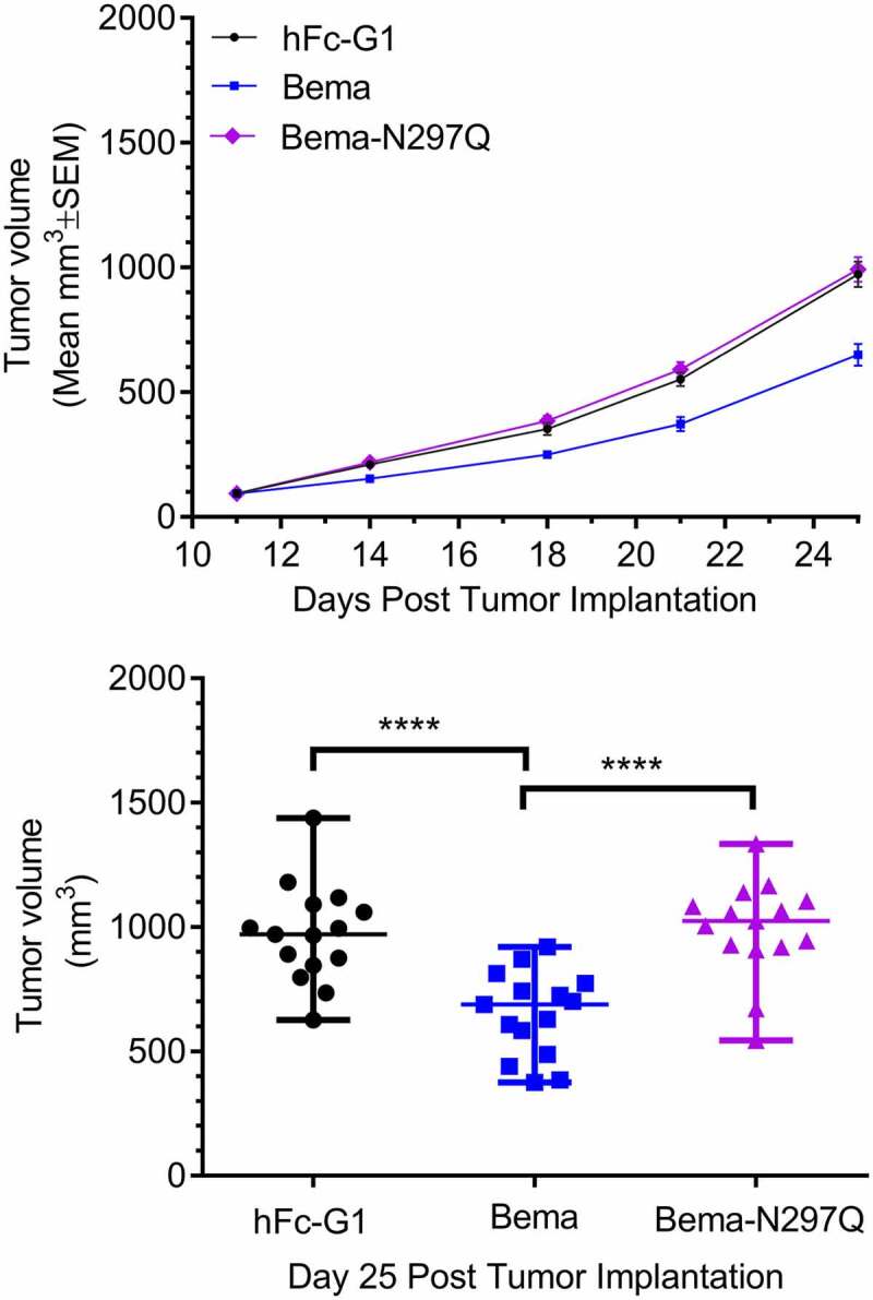
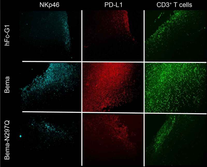
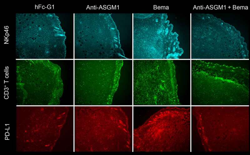
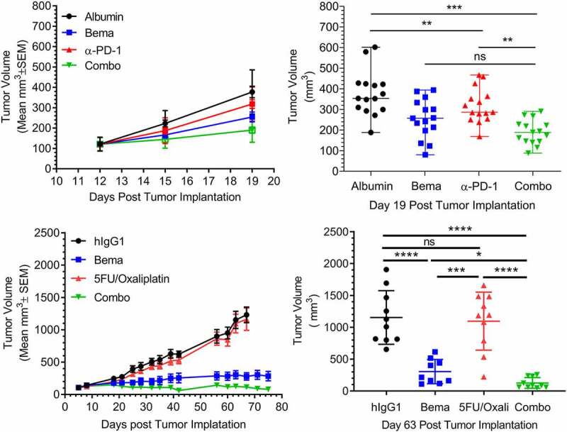
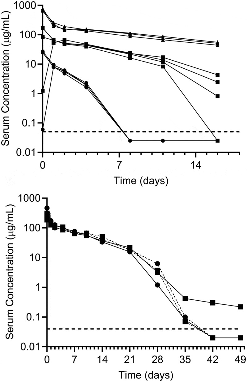
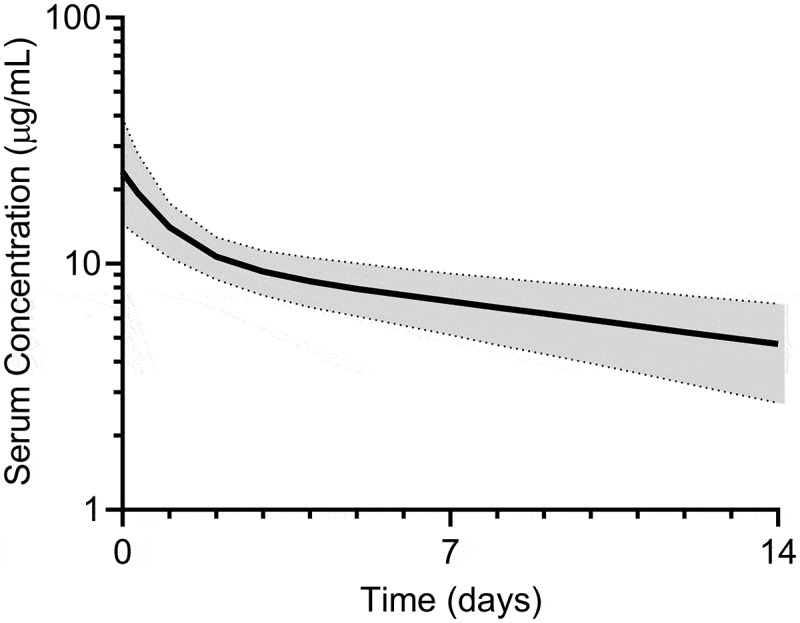
Bemarituzumab (FPA144) is a first-in-class, humanized, afucosylated immunoglobulin G1 monoclonal antibody (mAb) directed against fibroblast growth factor receptor 2b (FGFR2b) with two mechanisms of action against FGFR2b-overexpressing tumors: inhibition of FGFR2b signaling and enhanced antibody-dependent cell-mediated cytotoxicity (ADCC). Bemarituzumab is being developed as a cancer therapeutic, and we summarize here the key nonclinical data that supported moving it into clinical trials. Bemarituzumab displayed sub-nanomolar cross-species affinity for FGFR2b receptors, with >20-fold enhanced binding affinity to human Fc gamma receptor IIIa compared with the fucosylated version. In vitro, bemarituzumab induced potent ADCC against FGFR2b-expressing tumor cells, and inhibited FGFR2 phosphorylation and proliferation of SNU-16 gastric cancer cells in a concentration-dependent manner. In vivo, bemarituzumab inhibited tumor growth through inhibition of the FGFR2b pathway and/or ADCC in mouse models. Bemarituzumab demonstrated enhanced anti-tumor activity in combination with chemotherapy, and due to bemarituzumab-induced natural killer cell-dependent increase in programmed death-ligand 1, also resulted in enhanced anti-tumor activity when combined with an anti-programmed death-1 antibody. Repeat-dose toxicity studies established the highest non-severely-toxic dose at 1 and 100 mg/kg in rats and cynomolgus monkeys, respectively. In pharmacokinetic (PK) studies, bemarituzumab exposure increase was greater than dose-proportional, with the linear clearance in the expected dose range for a mAb. The PK data in cynomolgus monkeys were used to project bemarituzumab linear PK in humans, which were consistent with the observed human Phase 1 data. These key nonclinical studies facilitated the successful advancement of bemarituzumab into the clinic.

Keywords: Bemarituzumab; afucosylated antibody; antibody-dependent cell-mediated cytoxicity; phosphorylation in vitro; anti-FGFR2b antibody; anti-tumor efficacy; cell proliferation in vitro; fibroblast growth factor receptor; pharmacokinetics; toxicology.


Conflict of interest statement

This research was sponsored by FivePrime. All authors other than Rong Deng were employees of FivePrime and had a business and/or financial interest in the company. Rong Deng declare no potential conflicts of interest. Medical writing services were funded by FivePrime. Five Prime was acquired by Amgen Inc. on April 16th, 2021.

Figures

Figure 1.
Figure 1.
Bemarituzumab inhibits FGFR2 phosphorylation and FGFR2b-mediated proliferation of SNU-16 cells. a: FGFR2 phosphorylation (pFGFR2) in SNU-16 cells treated with varying concentrations of bemarituzumab followed by 100 ng/mL FGF7 is shown. FGFR2 phosphorylation was measured via ELISA. b: Proliferation of SNU-16 cells treated with varying concentrations of bemarituzumab followed by 100 ng/mL FGF7 is shown. Proliferation was measured via cell viability assay. The experiments were performed in triplicate; data are presented as mean ±standard error of mean (SEM) relative light units (RLU)
Figure 2.
Figure 2.
Bemarituzumab elicits potent induction of ADCC activity against FGFR2b-expressing cells. Specific lysis of target cells as a percentage of maximal lysis in the presence of varying concentrations of bemarituzumab or FPA144-F is shown: Ba/F3 cells expressing full-length human FGFR2b treated with bemarituzumab (Bema/FGFR2b); Ba/F3 cells expressing full-length human FGFR2b treated with FPA144-F (FPA144-F/GFGFR2b); Ba/F3 cells expressing full-length human FGFR2c treated with bemarituzumab (Bema/FGFR2c). Data are presented as mean ±standard error of mean (SEM); n = 3 per concentration/group
Figure 3.
Figure 3.
Bemarituzumab inhibits tumor growth in OCUM-2M tumor-bearing mice. Tumor volume (mm3) over time in OCUM-2M tumor-bearing mice treated with twice per week IV bemarituzumab at varying dose levels is shown. Albumin (5 mg/kg, IV) was used as the negative control. The first day of the treatment was day 4 post tumor implantation. Data are presented as mean ±standard error of mean (SEM); n = 15 per group
Figure 4.
Figure 4.
Bemarituzumab decreases FGFR2 signaling in SNU-16 tumor-bearing mice. Western blot analyses of pan-phosphorylated FGFR (phospho-FGFR), total FGFR2b, phosphorylated FRS2 (phospho-FRS2), and total FRS2 in tumors from SNU-16 tumor-bearing mice are shown. All results were compared to a common housekeeping enzyme (GAPDH). a: Tumors from mice treated with a single IV dose of bemarituzumab (10 mg/kg) were collected at 2, 6, 24, and 72 hours post treatment (n = 3 per time point). Control groups included tumors collected at 2 hours from naive animals (n = 2) and at 24 hours post IgG treatment (10 mg/kg; n = 3). Bemarituzumab decreased phospho-FGFR and phospho-FRS2 within 2 hours, and decreased total FGFR2b levels within 6 hours. b: Tumors from mice treated with twice weekly IP doses of 10 mg/kg bemarituzumab or albumin were collected at 82 days post first bemarituzumab/albumin treatment (n = 3 per group). Compared with the albumin control group, bemarituzumab decreased phospho-FGFR, phospho-FRS2, and total FGFR2b levels
Figure 5.
Figure 5.
Bemarituzumab, but not an ADCC-deficient FGFR2b antibody, leads to tumor suppression in a syngeneic tumor model with modest FGFR2b expression. a: Tumor volume (mm3) over time in mice bearing 4T1 mammary orthotopic tumors treated with twice per week bemarituzumab (10 mg/kg, IP) or bemarituzumab-N297Q (10 mg/kg, IP) are shown. Human Fc-IgG1 (hFc-G1,10 mg/kg, IP) was used as the negative control. The first day of treatment was Day 11 post tumor implantation. Data are presented as mean ±standard error of mean (SEM); n = 15 per group. b: at day 25 post tumor implantation, mean tumor volumes from mice in panel a were significantly different (****p < .0001). Statistical significance was determined by 1 way ANOVA followed by Tukey’s multiple comparisons test. Each symbol represents an individual animal
Figure 6.
Figure 6.
Bemarituzumab induces tumor-specific innate and adaptive immune profile changes. Mice bearing 4T1 tumors were treated with 20 mg/kg IP bemarituzumab (Bema), bemarituzumab-N297Q (Bema-N297Q), or human Fc-IgG1 (hFc-G1) on day 0 (when tumors reached approximately 150 mm3) and day 3, and then euthanized 24 hours later on day 4 for immunohistochemistry analysis. Representative fluorescence of NKp46, PD-L1, and CD3-positive T cells (CD3 T cells) 24 hours post second dose of treatment are shown
Figure 7.
Figure 7.
Bemarituzumab-induced increases in PD-L1 and CD3-positive T cells depends on NK cell recruitment. Mice bearing 4T1 tumors were treated with either: human Fc-IgG1 negative control (10 mg/kg, IP) on day 0 (when tumors reached approximately 125 mm3) and day 3 (hFc-G1), anti-ASGM1 (50 mg/kg, IV) once on day 0, bemarituzumab (10 mg/kg, IP) on day 0 and day 3 (Bema), or anti-ASGM1 (50 mg/kg, IV) on day 0 and bemarituzumab (10 mg/kg, IP) on day 0 and day 3 (anti-ASGM1 + Bema). Mice were euthanized on day 4 for immunohistochemistry analysis. Representative fluorescence of NKp46, PD-L1, and CD3-positive T cells (CD3+ T cells) are shown
Figure 8.
Figure 8.
Bemarituzumab displays enhanced anti-tumor activity when combined with anti-PD-1 or chemotherapeutics. a: Tumor volume (mm3) over time in mice bearing 4T1 mammary orthotopic tumors treated with twice per week bemarituzumab (10 mg/kg, IP), anti-PD-1 (5 mg/kg, IP), or bemarituzumab (10 mg/kg, IP) and anti-PD-1 (5 mg/kg, IP) in combination (Combo) are shown. Albumin (10 mg/kg, IP) was used as the negative control. The first day of the treatment was day 12 post tumor implantation. Data are presented as mean ±standard error of mean (SEM); n = 15 per group. b: at day 19 post tumor implantation, mean tumor volumes from mice in panel a were either statistically significant (**p < .01, ***p < .001) or not statistically significant (ns). Each symbol represents an individual animal. c: Tumor volume (mm3) over time in OCUM-2 M tumor-bearing mice treated with twice per week bemarituzumab (5 mg/kg, IP), weekly 5-FU (30 mg/kg, IP) and oxaliplatin (5 mg/kg, IP), or twice per week bemarituzumab (5 mg/kg, IP) and weekly 5-FU (30 mg/kg, IP)/oxaliplatin (5 mg/kg, IP) in combination (Combo) are shown. Human IgG1(5 mg/kg, IP) was used as the negative control (hIgG1). The first day of the treatment was post tumor implantation day 5 for 5-FU/oxaliplatin and day 11 for bemarituzumab. Data are presented as mean ±SEM; n = 9–10 per group. d: at day 63 post tumor implantation, mean tumor volumes from mice in Panel c were either statistically significant (*p < .05, ***p < .001, ****p < .0001) or not statistically significant (ns). Each symbol represents an individual animal. Statistical significance was determined by 1 way ANOVA followed by Tukey’s multiple comparisons
Figure 9.
Figure 9.
Bemarituzumab demonstrates biphasic disposition in rats and cynomolgus monkeys following a single IV injection. a: Bemarituzumab plasma concentration in rats following a single IV injection at 1.5 mg/kg (●; n = 4), 10 mg/kg (■; n = 4), or 30 mg/kg (▲; n = 3). Based on the concentration profiles, 2 animals (1 in the 1.5 mg/kg group and 1 in the 10 mg/kg group) were suspected of inadvertent subcutaneous dosing. b: Plasma concentrations of bemarituzumab and FPA144-F in cynomolgus monkeys following a single IV injection of bemarituzumab 10 mg/kg (–■̶; n = 1), bemarituzumab 13.5 mg/kg (–●̶; n = 1), or FPA144-F 10 mg/kg (-●-, -■-; n = 2). Each symbol represents a concentration time point for an individual animal. The lower limit of quantification (–) was 50 ng/mL
Figure 10.
Figure 10.
Projected human plasma concentration using PK data from cynomolgus monkeys. Predicted bemarituzumab plasma concentration-time profile (median [solid line], 5% and 95% quantiles [dashed lines], 90% prediction interval [shaded area]) in humans following 1 mg/kg IV administration. Bemarituzumab concentration-time profiles were scaled from cynomolgus monkey PK data using Dedrick approach with exponent of 0.85 and 1 for clearance and V1, respectively

References

    1. Korc M, Friesel RE.. The role of fibroblast growth factors in tumor growth. Curr Cancer Drug Targets. 2009;9(5):639–16. doi:10.2174/156800909789057006. - DOI - PMC - PubMed
    1. Miki T, Bottaro DP, Fleming TP, Smith CL, Burgess WH, Chan AM, Aaronson SA.. Determination of ligand-binding specificity by alternative splicing: two distinct growth factor receptors encoded by a single gene. Proc Natl Acad Sci USA. 1992;89(1):246–50. doi:10.1073/pnas.89.1.246. - DOI - PMC - PubMed
    1. Orr-Urtreger A, Bedford MT, Burakova T, Arman E, Zimmer Y, Yayon A, Givol D, Lonai P. Developmental localization of the splicing alternatives of fibroblast growth factor receptor-2 (FGFR2). Dev Biol. 1993;158(2):475–86. doi:10.1006/dbio.1993.1205. - DOI - PubMed
    1. Hattori Y, Odagiri H, Nakatani H, Miyagawa K, Naito K, Sakamoto H, Katoh O, Yoshida T, Sugimura T, Terada M. K-sam, an amplified gene in stomach cancer, is a member of the heparin-binding growth factor receptor genes. Proc Natl Acad Sci USA. 1990;87(15):5983–87. doi:10.1073/pnas.87.15.5983. - DOI - PMC - PubMed
    1. Jung E-J, Jung E-J, Min SY, Kim MA, Kim WH. Fibroblast growth factor receptor 2 gene amplification status and its clinicopathologic significance in gastric carcinoma. Hum Pathol. 2012;43(10):1559–66. doi:10.1016/j.humpath.2011.12.002. - DOI - PubMed

Publication types

MeSH terms

Substances