The Effect of Cognitive Load on the Retrieval of Long-Term Memory: An fMRI Study
- PMID: 34720904
- PMCID: PMC8548369
- DOI: 10.3389/fnhum.2021.700146
The Effect of Cognitive Load on the Retrieval of Long-Term Memory: An fMRI Study
Abstract
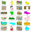
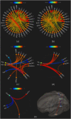
One of the less well-understood aspects of memory function is the mechanism by which the brain responds to an increasing load of memory, either during encoding or retrieval. Identifying the brain structures which manage this increasing cognitive demand would enhance our knowledge of human memory. Despite numerous studies about the effect of cognitive loads on working memory processes, whether these can be applied to long-term memory processes is unclear. We asked 32 healthy young volunteers to memorize all possible details of 24 images over a 12-day period ending 2 days before the fMRI scan. The images were of 12 categories relevant to daily events, with each category including a high and a low load image. Behavioral assessments on a separate group of participants (#22) provided the average loads of the images. The participants had to retrieve these previously memorized images during the fMRI scan in 15 s, with their eyes closed. We observed seven brain structures showing the highest activation with increasing load of the retrieved images, viz. parahippocampus, cerebellum, superior lateral occipital, fusiform and lingual gyri, precuneus, and posterior cingulate gyrus. Some structures showed reduced activation when retrieving higher load images, such as the anterior cingulate, insula, and supramarginal and postcentral gyri. The findings of this study revealed that the mechanism by which a difficult-to-retrieve memory is handled is mainly by elevating the activation of the responsible brain areas and not by getting other brain regions involved, which is a help to better understand the LTM retrieval process in the human brain.
Keywords: cognitive load; functional MRI (fMRI); long-term memory; memory retrieval; visual memory.
Copyright © 2021 Sisakhti, Sachdev and Batouli.
Conflict of interest statement
The authors declare that the research was conducted in the absence of any commercial or financial relationships that could be construed as a potential conflict of interest.
Figures


Similar articles
-
Emotional effect on cognitive control in implicit memory tasks in patients with schizophrenia.Neuroreport. 2015 Aug 5;26(11):647-55. doi: 10.1097/WNR.0000000000000405. Neuroreport. 2015. PMID: 26103120
-
Neural correlates of successful emotional episodic encoding and retrieval: An SDM meta-analysis of neuroimaging studies.Neuropsychologia. 2020 Jun;143:107495. doi: 10.1016/j.neuropsychologia.2020.107495. Epub 2020 May 13. Neuropsychologia. 2020. PMID: 32416099
-
Cognitive Functioning in Temporal Lobe Epilepsy: A BOLD-fMRI Study.Mol Neurobiol. 2017 Dec;54(10):8361-8369. doi: 10.1007/s12035-016-0298-0. Epub 2016 Dec 6. Mol Neurobiol. 2017. PMID: 27924527
-
An event-related fMRI study of the neural networks underlying the encoding, maintenance, and retrieval phase in a delayed-match-to-sample task.Brain Res Cogn Brain Res. 2005 May;23(2-3):207-20. doi: 10.1016/j.cogbrainres.2004.10.010. Epub 2004 Dec 9. Brain Res Cogn Brain Res. 2005. PMID: 15820629
-
Visual memory, visual imagery, and visual recognition of large field patterns by the human brain: functional anatomy by positron emission tomography.Cereb Cortex. 1995 Jan-Feb;5(1):79-93. doi: 10.1093/cercor/5.1.79. Cereb Cortex. 1995. PMID: 7719132 Clinical Trial.
Cited by
-
Investigation of the neural correlation with task performance and its effect on cognitive load level classification.PLoS One. 2023 Dec 21;18(12):e0291576. doi: 10.1371/journal.pone.0291576. eCollection 2023. PLoS One. 2023. PMID: 38127869 Free PMC article.
-
Understanding and Measuring the Cognitive Load of Amputees for Rehabilitation and Prosthesis Development.Arch Rehabil Res Clin Transl. 2022 Jul 13;4(3):100216. doi: 10.1016/j.arrct.2022.100216. eCollection 2022 Sep. Arch Rehabil Res Clin Transl. 2022. PMID: 36123983 Free PMC article. Review.
-
The subcortical brain regions influence the cortical areas during resting-state: an fMRI study.Front Hum Neurosci. 2024 Jun 26;18:1363125. doi: 10.3389/fnhum.2024.1363125. eCollection 2024. Front Hum Neurosci. 2024. PMID: 39055533 Free PMC article.
-
Eight brain structures mediate the age-related alterations of the working memory: forward and backward digit span tasks.Front Psychol. 2024 Sep 3;15:1377342. doi: 10.3389/fpsyg.2024.1377342. eCollection 2024. Front Psychol. 2024. PMID: 39295767 Free PMC article.
-
Strategies to improve the implementation of intensive lifestyle interventions for obesity.Front Public Health. 2023 Jul 25;11:1202545. doi: 10.3389/fpubh.2023.1202545. eCollection 2023. Front Public Health. 2023. PMID: 37559739 Free PMC article. No abstract available.
References
-
- Atkinson R., Holmgren J., Juola J. (1969). Processing time as influenced by the number of elements in a visual display. Percept. Psychophys. 6 321–326. 10.3758/BF03212784 - DOI
LinkOut - more resources
Full Text Sources

