. 2021 Oct 11:12:661217.
doi: 10.3389/fphar.2021.661217. eCollection 2021.

Investigation of Angiogenesis and Wound Healing Potential Mechanisms of Zinc Oxide Nanorods

Affiliations

Investigation of Angiogenesis and Wound Healing Potential Mechanisms of Zinc Oxide Nanorods

Amr Hassan et al. Front Pharmacol. .

Abstract

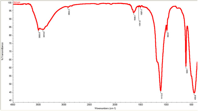
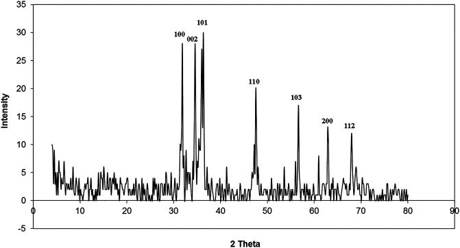
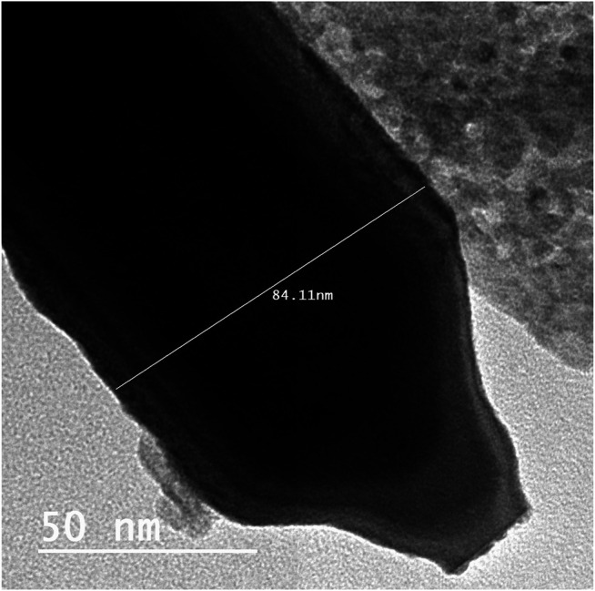
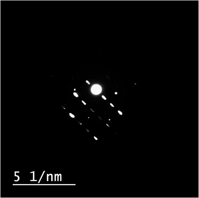
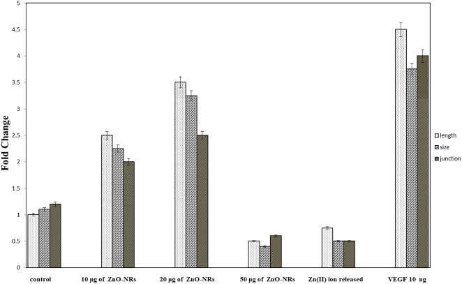
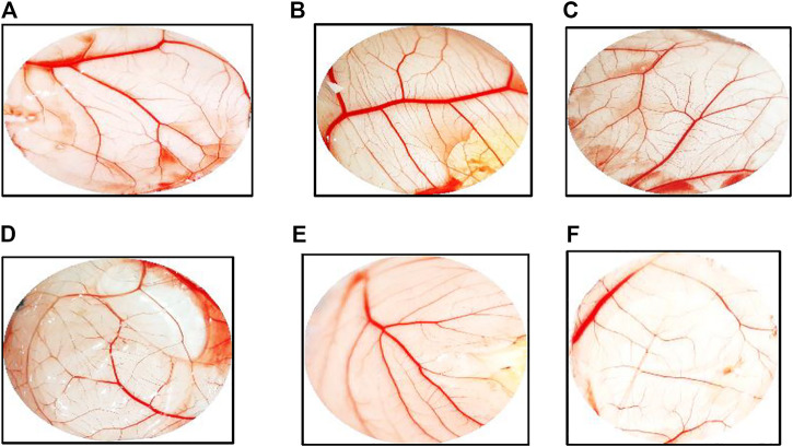
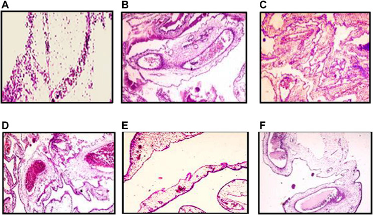
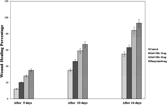
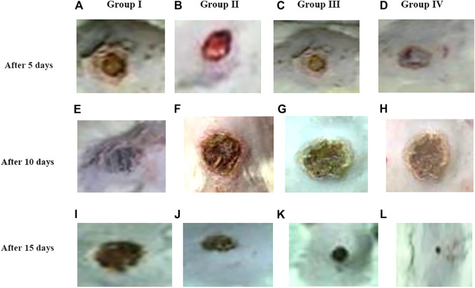
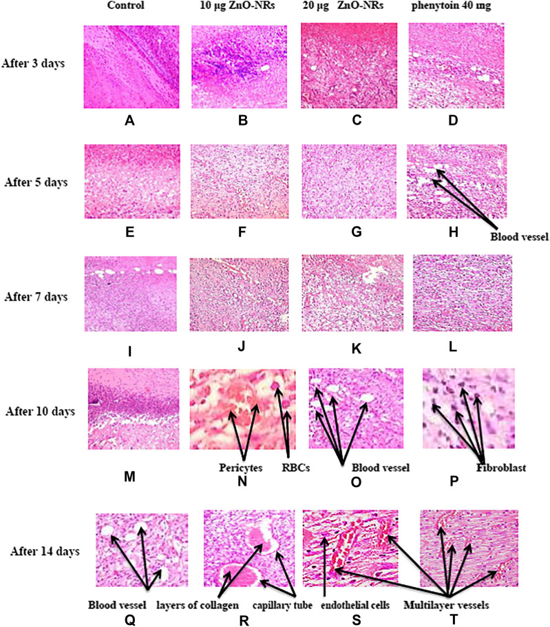
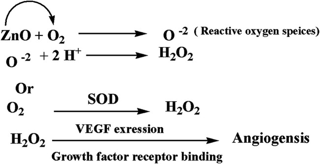
The angiogenesis process is an essential issue in tissue engineering. Zinc oxide nanorods are biocompatible metals capable of generating reactive oxygen species (ROS) that respond to induced angiogenesis through various mechanisms; however, released Zn (II) ions suppress the angiogenesis process. In this study, we fabricated green ZnO nanorods using albumin eggshell as a bio-template and investigate its angiogenic potential through chorioallantoic membrane assay and excision wound healing assay. This study demonstrated that angiogenesis and wound healing processes depend on pro-angiogenic factors as VEGF expression due to ZnO nanorods' exiting. Angiogenesis induced via zinc oxide nanorods may develop sophisticated materials to apply in the wound healing field.

Keywords: ROS; VEGF; angiogenesis; wound healing; zinc oxide nanorods.


Conflict of interest statement

The authors declare that the research was conducted in the absence of any commercial or financial relationships that could be construed as a potential conflict of interest.

Figures

FIGURE 1
FIGURE 1
FT-IR spectra ZnO nanorods (ZnO-NRs).
FIGURE 2
FIGURE 2
XRD patterns of ZnO nanorods (ZnO-NRs).
FIGURE 3
FIGURE 3
Transmission electron microscopy of ZnO nanorods (ZnO-NRs).
FIGURE 4
FIGURE 4
Diffraction index of ZnO nanorods (ZnO-NRs).
FIGURE 5
FIGURE 5
Released zinc (II) ions from ZnO nanorods by ICP-AES.
FIGURE 6
FIGURE 6
Quantitative real-time PCR measures mRNA levels of the vascular endothelial growth factor gene (VEGF).
FIGURE 7
FIGURE 7
Reactive oxygen species (ROS) of ZnO nanorods (ZnO-NRs) and released zinc (II) ions from ZnO nanorods (ZnO-NRs).
FIGURE 8
FIGURE 8
Glutathione peroxidase enzyme of ZnO nanorods (ZnO-NRs) and released zinc (II) ions from ZnO nanorods (ZnO-NRs).
FIGURE 9
FIGURE 9
Angiogenesis parameter quantitative of ZnO nanorods (ZnO-NRs) and released zinc (II) ions using chicken chorioallantoic membrane (CAM) assay.
FIGURE 10
FIGURE 10
Chicken chorioallantoic membrane (CAM) assay. (A) Negative control (non-treated). (B) CAM treated with 10 μg/ml of ZnO nanorods (ZnO-NRs). (C) CAM treated with 20 μg/ml of ZnO nanorods (ZnO-NRs). (D) CAM treated with 10 ng of positive control VEGF. (E) CAM treated with 50 μg/ml of ZnO nanorods (ZnO-NRs). (F) CAM treated with released Zn (II) ions.
FIGURE 11
FIGURE 11
Histopathology pattern of chick embryo chorioallantoic (CAM) assay. (A) Negative control (non-treated). (B) CAM treated with 10 μg/ml of ZnO nanorods (ZnO-NRs). (C) CAM treated with 20 μg/ml of ZnO nanorods (ZnO-NRs). (D) CAM treated with 10 ng of positive control VEGF. (E) CAM treated with 50 μg/ml of ZnO nanorods (ZnO-NRs). (F) CAM treated with released Zn (II) ions.
FIGURE 12
FIGURE 12
Wound healing percentages after remediation with ZnO nanorods (ZnO-NRs), phenytoin compared with control (non-treated).
FIGURE 13
FIGURE 13
Morphology of wound healing after exposure with ZnO nanorods (ZnO-NRs), phenytoin compared with control (non-treated). (A) Negative control (non-treated) after 5 days. (B) Wound healing after exposure with 10 μg/ml of ZnO nanorods (ZnO-NRs) after 5 days. (C) Wound healing after exposure with 20 μg/ml of ZnO nanorods (ZnO-NRs) after 5 days. (D) Wound healing after exposure with phenytoin as positive control after 5 days. (E) Negative control (non-treated) after 10 days. (F) Wound healing after exposure with 10 μg/ml of ZnO nanorods (ZnO-NRs) after 10 days. (G) Wound healing after exposure with 20 μg/ml of ZnO nanorods (ZnO-NRs) after 10 days. (H) Wound healing after exposure with phenytoin as positive control after 10 days. (I) Negative control (non-treated) after 15 days. (J) Wound healing after exposure with 10 μg/ml of ZnO nanorods (ZnO-NRs) after 15 days. (K) Wound healing after exposure with 20 μg/ml of ZnO nanorods (ZnO-NRs) after 15 days. (L) Wound healing after exposure with phenytoin as positive control after 15 days
FIGURE 14
FIGURE 14
Histopathology pattern of wound healing after treatment with ZnO nanorods (ZnO-NRs), phenytoin compared with control (non-treated). (A) Negative control (non-treated) after 3 days. (B) Wound healing after treatment with 10 μg/ml of ZnO nanorods (ZnO-NRs) after 3 days. (C) Wound healing after treatment with 20 μg/ml of ZnO nanorods (ZnO-NRs) after 3 days. (D) Wound healing after treatment with phenytoin as positive control after 3 days. (E) Negative control (non-treated) after 5 days. (F) Wound healing after treatment with 10 μg/ml of ZnO nanorods (ZnO-NRs) after 5 days. (G) Wound healing after treatment with 20 μg/ml of ZnO nanorods (ZnO-NRs) after 5 days. (H) Wound healing after treatment with phenytoin as positive control after 5 days. (I) Negative control (non-treated) after 7 days. (J) Wound healing after treatment with 10 μg/ml of ZnO nanorods (ZnO-NRs) after 7 days. (K) Wound healing after treatment with 20 μg/ml of ZnO nanorods (ZnO-NRs) after 7 days. (L) Wound healing after treatment with phenytoin as positive control after 7 days. (M) Negative control (non-treated) after 10 days. (N) Wound healing after treatment with 10 μg/ml of ZnO nanorods (ZnO-NRs) after 10 days. (O) Wound healing after treatment with 20 μg/ml of ZnO nanorods (ZnO-NRs) after 10 days. (P) Wound healing after treatment with phenytoin as positive control after 10 days. (Q) Negative control (non-treated) after 14 days. (R) Wound healing after treatment with 10 μg/ml of ZnO nanorods (ZnO-NRs) after 14 days. (S) Wound healing after treatment with 20 μg/ml of ZnO nanorods (ZnO-NRs) after 14 days. (T) Wound healing after treatment with phenytoin as positive control after 14 days.
FIGURE 15
FIGURE 15
A plausible mechanism of angiogenesis

References

    1. Augustine R., Dominic E. A., Reju I., Kaimal B., Kalarikkal N., Thomas S. (2014). Investigation of Angiogenesis and its Mechanism Using Zinc Oxide Nanoparticle-Loaded Electrospun Tissue Engineering Scaffolds. RSC Advances 93, 51528–51536. 10.1039/C4RA07361D - DOI
    1. Ausprunk D. H., Knighton D. R., Folkman J. (1975). Vascularization of Normal and Neoplastic Tissues Grafted to the Chick Chorioallantois. Role of Host and Preexisting Graft Blood Vessels. Am. J. Pathol., 79 (3), 597, 618. - PMC - PubMed
    1. Bancroft J. D., Stevans A., Turner D. R. (2013). Theory and Practice of Histological Techniques. 4th Ed. Edinburgh, London, Melbourne, New York: Churchill Livingstone.
    1. Bao S.-J., Lei C., Xu M.-W., Cai C.-J., Jia D.-Z. (2012). Environment-friendly Biomimetic Synthesis of TiO2nanomaterials for Photocatalytic Application. Nanotechnology, 23 (20), 205601. 10.1088/0957-4484/23/20/205601 - DOI - PubMed
    1. Barui A. K., Veeriah V., Mukherjee S., Manna J., Patel A. K., Patra S., et al. (2012). Zinc Oxide Nanoflowers Make New Blood Vessels. Nanoscale 4 (24), 7861–7869. 10.1039/c2nr32369a - DOI - PubMed

LinkOut - more resources