. 2021 Oct 14:12:742848.
doi: 10.3389/fimmu.2021.742848. eCollection 2021.

21-Hydroxylase-Specific CD8+ T Cells in Autoimmune Addison's Disease Are Restricted by HLA-A2 and HLA-C7 Molecules

Affiliations

21-Hydroxylase-Specific CD8+ T Cells in Autoimmune Addison's Disease Are Restricted by HLA-A2 and HLA-C7 Molecules

Alexander Hellesen et al. Front Immunol. .

Abstract

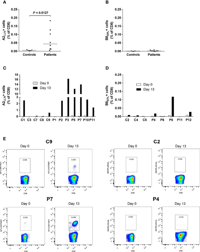
Objectives: CD8+ T cells targeting 21-hydroxylase (21OH) are presumed to play a central role in the destruction of adrenocortical cells in autoimmune Addison's disease (AAD). Earlier reports have suggested two immunodominant CD8+ T cell epitopes within 21OH: LLNATIAEV (21OH342-350), restricted by HLA-A2, and EPLARLEL (21OH431-438), restricted by HLA-B8. We aimed to characterize polyclonal CD8+ T cell responses to the proposed epitopes in a larger patient cohort with AAD.

Methods: Recombinant fluorescent HLA-peptide multimer reagents were used to quantify antigen-specific CD8+ T cells by flow cytometry. Interferon-gamma (IFNγ) Elispot and biochemical assays were used to functionally investigate the 21OH-specific T cells, and to map the exactly defined epitopes of 21OH.

Results: We found a significantly higher frequency of HLA-A2 restricted LLNATIAEV-specific cells in patients with AAD than in controls. These cells could also be expanded in vitro in an antigen specific manner and displayed a robust antigen-specific IFNγ production. In contrast, only negligible frequencies of EPLARLEL-specific T cells were detected in both patients and controls with limited IFNγ response. However, significant IFNγ production was observed in response to a longer peptide encompassing EPLARLEL, 21OH430-447, suggesting alternative dominant epitopes. Accordingly, we discovered that the slightly offset ARLELFVVL (21OH434-442) peptide is a novel dominant epitope restricted by HLA-C7 and not by HLA-B8 as initially postulated.

Conclusion: We have identified two dominant 21OH epitopes targeted by CD8+ T cells in AAD, restricted by HLA-A2 and HLA-C7, respectively. To our knowledge, this is the first HLA-C7 restricted epitope described for an autoimmune disease.

Keywords: 21-hydroxylase; Addison’s disease; CD8+ T cells; autoimmune; epitopes.


Conflict of interest statement

The authors declare that the research was conducted in the absence of any commercial or financial relationships that could be construed as a potential conflict of interest.

Figures

Figure 1
Figure 1
Increased frequencies of LLNATIAEV-specific CD8+T cells in AAD patients. (A) PBMCs from HLA-A2 positive AAD patients (n = 8) and controls (n = 5) were stained with A2*LLNATIAEV dextramers ex vivo and analyzed with flow cytometry. (B) PBMCs from HLA-B8 positive AAD patients (n = 8) and controls (n = 4) were stained with B8*EPLARLEL dextramers ex vivo and analyzed with flow cytometry. (C) PBMCs from HLA-A2 AAD patients (n = 7) and controls (n = 5) were stained with A2*LLNATIAEV dextramers following 13 days of peptide-induced in vitro expansion and analyzed with flow cytometry. Differences were not statistically significant. (D) PBMCs from HLA-B8 AAD patients (n = 5) and controls (n = 3) were stained with B8*EPLARLEL dextramers following 13 days of peptide-induced in vitro expansion and analyzed with flow cytometry. The graphs (A–D) show the frequencies of dextramer-binding CD8+ T cells. Lines (A, B) represent median values. Statistical analyses were performed using Mann-Whitney U-test; P-values <0.05 are shown. (E) Representative flow cytometry dextramer plots from two HLA-A2 (C9, P7) and two HLA-B8 (C2, P4) individuals ex vivo and following expansion (day 13). A strict, fixed gate was used to distinguish dextramer-positive from negative cells. Gate numbers represent the frequencies (percentage) of dextramer-positive cells among total CD8+ T cells. Statistical analyses were performed using Mann-Whitney U-test; P-values <0.05 are shown.
Figure 2
Figure 2
Increased IFNγ responses to LLNATIAEV, but not EPLARLEL, in patients with AAD. (A) PBMCs from HLA-A2 positive individuals were stimulated with LLNATIAEV peptide ex vivo (AAD patients n = 8, controls n = 5) or restimulated following 13 days of peptide-induced in vitro expansion (AAD patients n = 7, controls n = 4). (B) PBMCs from HLA-B8 positive individuals were stimulated with EPLARLEL peptide ex vivo (AAD patients n = 5, controls n = 3) or restimulated following 13 days of peptide-induced in vitro expansion (AAD patients n = 5, controls n = 3). The number of IFNγ-secreting cells (SFC) was measured by ELISPOT and is expressed as SFC per 6x10⁵ PBMCs. Lines represent median values. Statistical analyses were performed using Mann-Whitney U-test; P-values <0.05 are shown.
Figure 3
Figure 3
Fine-mapping of T cell responses towards Pep34 (21OH430-447 GEPLARLELFVVLTRLLQ). PBMCs from patients (n = 10) and controls (n =5) were stimulated ex vivo with individual overlapping 9-mer (Pep1-10) peptides covering Pep34, as well as Pep34 itself, and subjected to IFNγ ELISPOT. Of note, Pep2 represents EPLARLELF. Results are expressed as IFNγ SFCs per 6x10⁵ PBMCs and lines represent median values.
Figure 4
Figure 4
IFNγ producing ARLELFVVL-specific CD8+ T cells are enriched in AAD patients. (A) PBMCs from HLA-C7-positive AAD patients (n = 13) and controls (n = 14) were stained with C7*ARLELFVVL streptamers ex vivo and analyzed with flow cytometry. (B) PBMC from HLA-C7-positive AAD patients (n = 6) and controls (n = 5) were stimulated with ARLELFVVL peptide and expanded for 13 days in vitro, before being stained with C7*ARLELFVVL streptamers and analyzed with flow cytometry. The graphs (A, B) show the frequencies of streptamer-binding CD8+ T cells. (C) Examples of flow cytometry streptamer plots from HLA-C7 individuals (C28, P19) ex vivo and following expansion (day 13). A strict, fixed gate was used to distinguish streptamer-positive from negative cells. Gate numbers represent the frequencies (percentage) of streptamer-positive cells among total CD8+ T cells. (D) PBMCs from AAD patients (n =14) and controls (n = 14) were stimulated with ARLELFVVL or Pep34 ex vivo to detect IFNγ-SFCs. The amount was measured by ELISPOT and is expressed as SFCs per 6x10⁵ PBMCs after background subtraction. Lines represent median values (A, D). Statistical analyses were performed using Mann-Whitney U-test; P-values <0.05 are shown, * in (B) signifies a statistically significant difference of streptamer-positive cells between patients and controls on day 13 (P = 0.0043).

References

    1. Husebye ES, Anderson MS, Kampe O. Autoimmune Polyendocrine Syndromes. N Engl J Med (2018) 378(12):1132–41. doi: 10.1056/NEJMra1713301 - DOI - PMC - PubMed
    1. Erichsen MM, Lovas K, Skinningsrud B, Wolff AB, Undlien DE, Svartberg J, et al. . Clinical, Immunological, and Genetic Features of Autoimmune Primary Adrenal Insufficiency: Observations From a Norwegian Registry. J Clin Endocrinol Metab (2009) 94(12):4882–90. doi: 10.1210/jc.2009-1368 - DOI - PubMed
    1. Betterle C, Dal Pra C, Mantero F, Zanchetta R. Autoimmune Adrenal Insufficiency and Autoimmune Polyendocrine Syndromes: Autoantibodies, Autoantigens, and Their Applicability in Diagnosis and Disease Prediction. Endocr Rev (2002) 23(3):327–64. doi: 10.1210/edrv.23.3.0466 - DOI - PubMed
    1. Coco G, Dal Pra C, Presotto F, Albergoni MP, Canova C, Pedini B, et al. . Estimated Risk for Developing Autoimmune Addison's Disease in Patients With Adrenal Cortex Autoantibodies. J Clin Endocrinol Metab (2006) 91(5):1637–45. doi: 10.1210/jc.2005-0860 - DOI - PubMed
    1. Laureti S, De Bellis A, Muccitelli VI, Calcinaro F, Bizzarro A, Rossi R, et al. . Levels of Adrenocortical Autoantibodies Correlate With the Degree of Adrenal Dysfunction in Subjects With Preclinical Addison's Disease. J Clin Endocrinol Metab (1998) 83(10):3507–11. doi: 10.1210/jc.83.10.3507 - DOI - PubMed

Publication types

MeSH terms