

doi: 10.3897/CompCytogen.v15.i4.64350.
eCollection 2021.
Study of chromatin diminution in Cyclopskolensis (Copepoda, Crustacea) by radiobiological methods
Affiliations
- PMID: 34721816
- PMCID: PMC8516822
- DOI: 10.3897/CompCytogen.v15.i4.64350
Item in Clipboard
Study of chromatin diminution in Cyclopskolensis (Copepoda, Crustacea) by radiobiological methods
Comp Cytogenet.
.
Abstract
The experimental results show that at doses of 20 Gy and 100 Gy, the development of Cyclopskolensis Lilljeborg, 1901 (Copepoda, Cyclopoida) embryos ceases at the 16-cell stage, without affecting the course of chromatin diminution. A dose of 200 Gy terminated the process of chromatin diminution in some of the embryos. These results support the hypothesis that cytoplasmic factors in the egg play an important role in the process of chromatin diminution.
Keywords: Copepoda; embryogenesis; radiation.
Andrey Grishanin, Oksana Chinyakova.
Figures

Anaphase of 3rd cleavage division of C.kolensis embryo at 5 Gy (dose of ionizing radiation). Designations: Chr- chromosome, Br- bridge, Fr- fragment.

Metaphase of a C.kolensis embryo cell after irradiation at a dose of 100 Gy. Chr- chromosome, Fr- fragment.
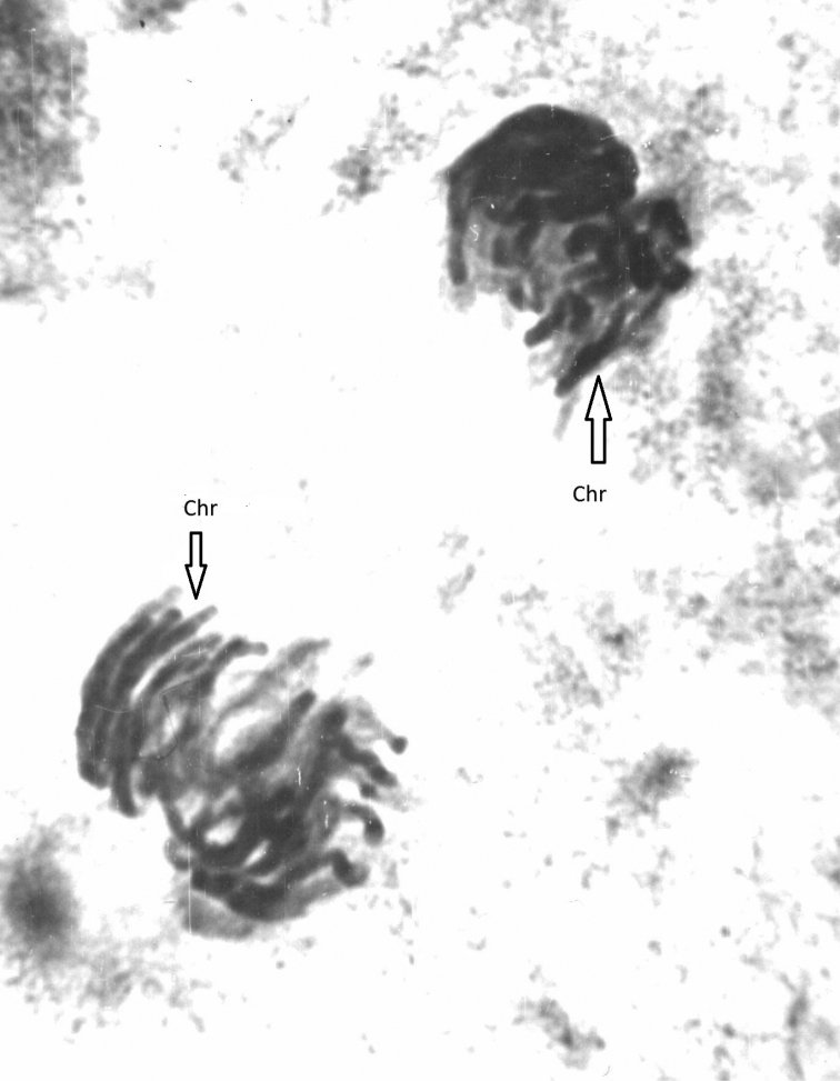
Anaphase of 3-d cleavage division of C.kolensis embryo in the control experiment. Chr- chromosome.

The percentage of Cyclopskolensis embryos that reached a particular cell stage of embryonic development in embryos exposed to different levels of radiation for 24 hours.
Similar articles
-
Chromatin diminution as a tool to study some biological problems.Comp Cytogenet. 2024 Feb 8;18:27-49. doi: 10.3897/compcytogen.17.112152. eCollection 2024. Comp Cytogenet. 2024. PMID: 38369988 Free PMC article.
-
Chromatin diminution in Copepoda (Crustacea): pattern, biological role and evolutionary aspects.Comp Cytogenet. 2014 Jan 9;8(1):1-10. doi: 10.3897/CompCytogen.v8i1.5913. eCollection 2014. Comp Cytogenet. 2014. PMID: 24744830 Free PMC article.
-
Chromatin Diminution in Cyclops kolensis Lill. (Copepoda, Crustacea) as a Radical Way to Inactivate Redundant Genome in Somatic Cells.Cytogenet Genome Res. 2018;156(3):165-172. doi: 10.1159/000494157. Epub 2018 Oct 31. Cytogenet Genome Res. 2018. PMID: 30376670 Review.
-
[The peculiarities of the radiation mutagenesis of Cyclops kolensis and Cyclops insignis (Crustacea, Copepoda)].Radiats Biol Radioecol. 2005 May-Jun;45(3):294-8. Radiats Biol Radioecol. 2005. PMID: 16080619 Russian.
-
[Chromatin diminution at the border of the XX and XXI centuries].Tsitologiia. 2006;48(5):379-97. Tsitologiia. 2006. PMID: 16892848 Review. Russian.
Cited by
-
Chromatin diminution as a tool to study some biological problems.Comp Cytogenet. 2024 Feb 8;18:27-49. doi: 10.3897/compcytogen.17.112152. eCollection 2024. Comp Cytogenet. 2024. PMID: 38369988 Free PMC article.
References
-
- Amma K. (1911) Uber die differenzeirung der Keimbahnzellen bei den Copepoden. Archive Experimental Zellforschung 6: 497–576.
-
- Boveri Th. (1910) Die Potenzen der Ascaris-Blastomeren bei abgeanderter Furchung Zugleich ein Beitrag zur Frage quilitatve ungleicher Chromosomenteilung. Festschrift für R. Hertwig, Jena 3: 131–214.
-
- Grishanin AK. (1995) Comparative electron microscopic study of chromosomes and interphase nuclei in cells of Cyclopskolensis (Copepoda, Crustacea) before and after chromatin diminution. Russian Journal of Developmental Biology 26(3): 153–158.
LinkOut - more resources
Full Text Sources