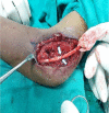
A missed medial humeral epicondyle fracture with incarcerated fragment in the elbow joint and ulnar nerve palsy: A rare case report
- PMID: 34721854
- PMCID: PMC8543049
- DOI: 10.1002/ccr3.4982
A missed medial humeral epicondyle fracture with incarcerated fragment in the elbow joint and ulnar nerve palsy: A rare case report
Abstract
Medial epicondyle fracture associated with incarcerated intra-articular fragment and ulnar nerve palsy is uncommon and frequently missed. We report a case of 13-year-old boy with incarcerated medial epicondyle fracture fragment in ulnohumeral joint and ulnar nerve palsy, which was managed successfully by open reduction internal fixation and ulnar nerve transposition.
Keywords: elbow joint; incarcerated; medial epicondyle fracture; ulnar nerve palsy; ulnohumeral joint.
© 2021 The Authors. Clinical Case Reports published by John Wiley & Sons Ltd.
Conflict of interest statement
None.
Figures


Similar articles
-
Intra-articular Entrapment of Medial Epicondyle Fracture Fragment in Elbow Joint Dislocation Causing Ulnar Neuropraxia: A Case Report.Malays Orthop J. 2017 Mar;11(1):82-84. doi: 10.5704/MOJ.1703.016. Malays Orthop J. 2017. PMID: 28435584 Free PMC article.
-
Medial Humeral Epicondyle Fracture Incarcerated Into the Elbow Joint in an Adolescent Patient With Ulnar Nerve Palsy.Cureus. 2023 Feb 1;15(2):e34502. doi: 10.7759/cureus.34502. eCollection 2023 Feb. Cureus. 2023. PMID: 36874314 Free PMC article.
-
Open Reduction Internal Fixation of a Medial Epicondyle Avulsion Fracture With Incarcerated Fragment.J Orthop Trauma. 2019 Aug;33 Suppl 1:S9-S10. doi: 10.1097/BOT.0000000000001530. J Orthop Trauma. 2019. PMID: 31290819
-
Delayed Reconstruction Following Incarceration of the Medial Epicondyle in the Elbow Joint: A Case Report and Review of the Literature.JBJS Case Connect. 2018 Jul-Sep;8(3):e69. doi: 10.2106/JBJS.CC.17.00239. JBJS Case Connect. 2018. PMID: 30211712 Review.
-
[Incarcerated epitrochlear fracture with a cubital nerve injury].Rev Esp Cir Ortop Traumatol. 2013 Sep-Oct;57(5):375-8. doi: 10.1016/j.recot.2013.06.002. Epub 2013 Sep 20. Rev Esp Cir Ortop Traumatol. 2013. PMID: 24071050 Review. Spanish.
References
-
- Gottschalk HP, Eisner E, Hosalkar HS. Medial epicondyle fractures in the pediatric population. J Am Acad Orthop Surg. 2012;20(4):223‐232. - PubMed
-
- Ramsey RW, Herman MJ. Medial epicondyle fractures. In: Abzug JM, Herman MJ, eds. Pediatric Elbow Fractures. Springer. 2018;95‐109.
-
- Lima S, Correia JF, Ribeiro RP, et al. A rare case of elbow dislocation associated with unrecognized fracture of medial epicondyle and delayed ulnar neuropathy in pediatric age. J Shoulder Elbow Surg. 2013;22(3):e9‐e11. - PubMed
-
- Haflah NHM, Ibrahim S, Sapuan J, Abdullah S. An elbow dislocation in a child with missed medial epicondyle fracture and late ulnar nerve palsy. J Pediatr Orthop B. 2010;19(5):459‐461. - PubMed
Publication types
LinkOut - more resources
Full Text Sources

