. 2021 Oct 14:9:723801.
doi: 10.3389/fcell.2021.723801. eCollection 2021.

Iron-Dependent Autophagic Cell Death Induced by Radiation in MDA-MB-231 Breast Cancer Cells

Affiliations

Iron-Dependent Autophagic Cell Death Induced by Radiation in MDA-MB-231 Breast Cancer Cells

Shumei Ma et al. Front Cell Dev Biol. .

Abstract

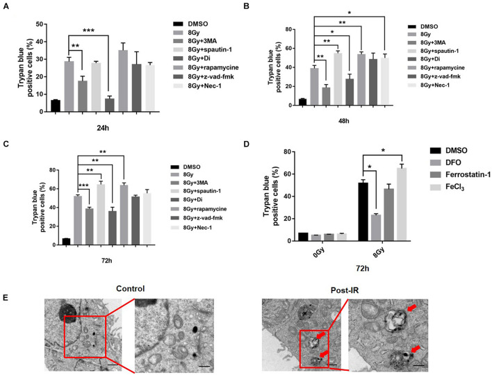
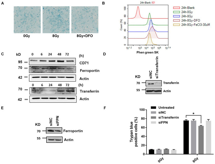
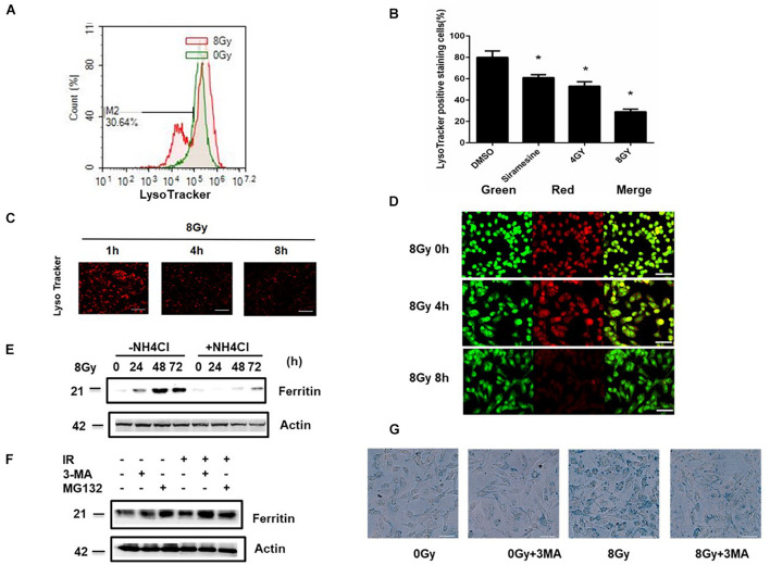
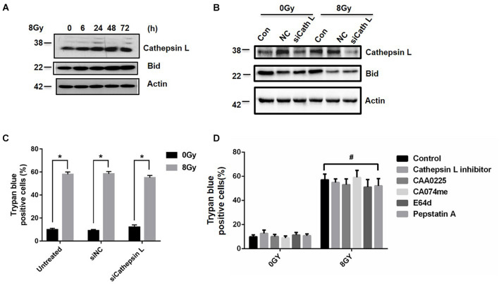
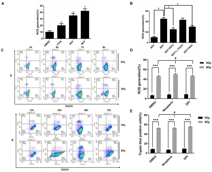
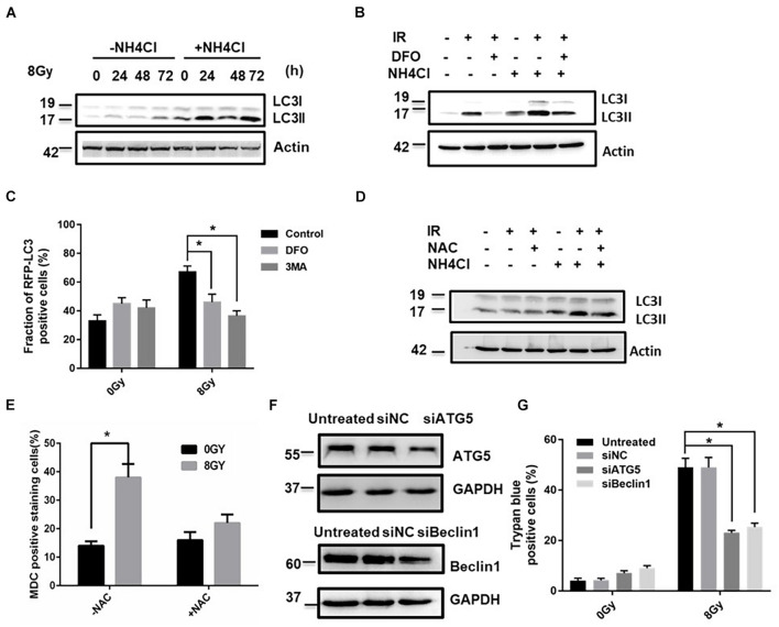
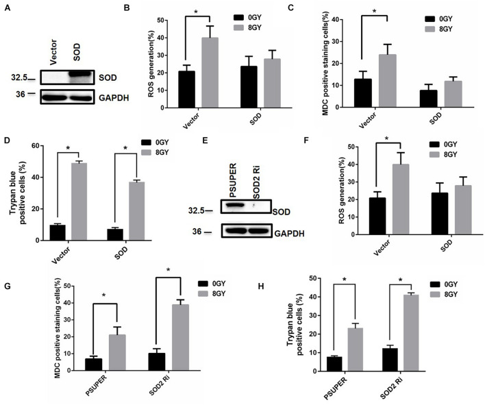
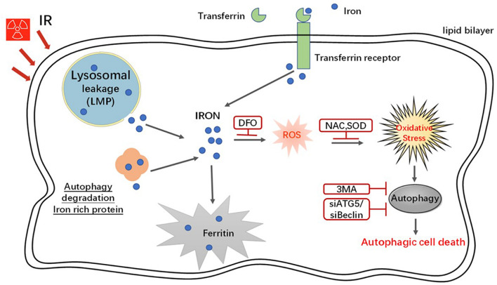
In radiation oncology, ionizing radiation is used to kill cancer cells, in other words, the induction of different types of cell death. To investigate this cellular death and the associated iron accumulation, the transfer, release, and participation of iron after radiation treatment was analyzed. We found that radiation-induced cell death varied in different breast cancer cells and autophagy was induced in MDA-MB-231 and BT549 cells (triple negative breast cancer cell line) rather than in MCF-7 and zr-75 cells. Iron chelator deferoxamine (DFO), the autophagy inhibitor 3MA, silencing of the autophagy-related genes ATG5, and Beclin 1 could decrease radiation induced cell death in MDA-MB-231 cells, while inhibitors of apoptosis such as Z-VAD-FMK, ferroptosis inhibitor ferrostatin-1 (Fer-1), and necroptosis inhibitor Necrostatin-1 showed no change. This suggests the occurrence of autophagic cell death. Furthermore, we found that iron accumulation and iron regulatory proteins, including transferrin (Tf), transferrin receptor (CD71), and Ferritin (FTH), increased after radiation treatment, and the silencing of transferrin decreased radiation-induced cell death. In addition, radiation increased lysosomal membrane permeabilization (LMP) and the release of lysosomal iron and cathepsins, while cathepsins silencing failed to change cell viability. Radiation-induced iron accumulation increased Reactive oxygen species (ROS) generation via the Fenton reaction and increased autophagy in a time-dependent manner. DFO, N-acetylcysteine (NAC), and overexpression of superoxide dismutase 2 (SOD2) decreased ROS generation, autophagy, and cell death. To summarize, for the first time, we found that radiation-induced autophagic cell death was iron-dependent in breast cancer MDA-MB-231 cells. These results provide new insights into the cell death process of cancers and might conduce to the development and application of novel therapeutic strategies for patients with apoptosis-resistant breast cancer.

Keywords: autophagy; breast cancer; iron; lysosome membrane permeabilization (LMP); radiation; reactive oxygen species (ROS).


Conflict of interest statement

The authors declare that the research was conducted in the absence of any commercial or financial relationships that could be construed as a potential conflict of interest.

Figures

FIGURE 1
FIGURE 1
Radiation induced cell death in MDA-MB-231 breast cancer cells. MDA-MB-231 were pretreated with different inhibitors of cell death, i.e., 3MA (2 mM), spautin-1 (3 μM), rapamycin (5 nM), z-VAD-fmk (10 μM), Necrostain-1 (10 μM), Digitoxigenin (Di, 5 μM) and cell deaths were quantified at 24, 48, 72 h after radiation (A–C). Cells were pretreated with FeCl3 (30 μM, pretreated for 3 h), DFO (0.1 mM), Ferrostain-1 (5 μm) for 1 h, cell death was quantified at 48 h after 8 Gy radiation (D). Electron microscopy analysis of autophagosome in the MDA-MB-231 cells was made after radiation treatment. Red arrow pointed to autophagosomes (E). These results were representative of three independent experiments and expressed as means ± SD. P-values were calculated using repeated measure ANOVA with P < 0.05 being considered as statistical significance, *P < 0.05, **P < 0.01, and ***P < 0.001.
FIGURE 2
FIGURE 2
Radiation increased iron level and ROS generation in MDA-MB-231 breast cancer cells. Cells were pretreated with DFO, followed by 8 Gy radiation, Prussian blue staining for intracellular iron was performed (A). Cells were pretreated with DFO or FeCl3 before 8 Gy radiation. Intracellular chelatable iron was detected using the fluorescent indicator Phen green SK with Flow cytometry (B). The expressions of iron-related proteins Transferrin (Tf), Transferrin receptor (CD71), and Ferroportin (FPN) were performed after 8 Gy treatment (C). Knockdown of Transferrin, FPN by siRNA as demonstrated by western blot (D,E). Cells were transfected with control siRNA (siNC) and siRNA against Transferrin (si Tf), Ferroprotein (siFPN) for 48 h, the amount of cell death was determined at 48 h after 8 Gy radiation (F). These results were representative of three independent experiments and expressed as means ± SD. P-values were calculated using repeated measure ANOVA with P < 0.05 being considered as statistical significance, *P < 0.05.
FIGURE 3
FIGURE 3
Radiation induced Lysosomal membrane permeabilization (LMP) in MDA-MB-231 breast cancer cells. MDA-MB-231 cells were treated with 4 Gy, 8 Gy radiation respectively, the amount of LMP was detected at 4, 8 h with LysoTracker and analyzed by flow cytometry (A–C) or with AO staining (D). In the presence or absence of NH4Cl, the Ferritin (FTH) expression were detected after 8 Gy treatment at 24 h (E). Cells were pretreated with 3MA (2 mM) or MG132 (1 μM) for 1 h before 8 Gy radiation, the Ferritin (FTH) expression were detected at 24 h (F). Cells were pretreated with 3MA before 8 Gy radiation, the intracellular iron was evaluated with Prussian blue staining at 24 h by light microscopy (G). These results were representative of three independent experiments and expressed as means ± SD. P-values were calculated using repeated measure ANOVA with P < 0.05 being considered as statistical significance, *P < 0.05.
FIGURE 4
FIGURE 4
Inhibition of Cathepsin L fails to block radiation-induced cell death. Cathepsin L expression was analyzed by western blot in MDA-MB-231 cells at 6, 24, 48, 72 h after 8 Gy radiation (A). After the knockdown of Cathepsin L by siRNAs, Cathepsin L and Bid expressions were determined after 8 Gy radiation (B). Cells were untransfected or transfected with control siRNA (siNC) and siRNA against Cathepsin L (siCathepsin L) for 48 h before 8 Gy radiation, then cell death was determined (C). Cells were pretreated with CAA0225 (a Cathepsin L inhibitor, 10 μg/ml) for 1 h before 8 Gy treatment, cathepsin L and Cleaved-Caspase3 expression were tested (D). These results were representative of three independent experiments and expressed as means ± SD. P-values were calculated using repeated measure ANOVA with P < 0.05 being considered as statistical significance, *P < 0.05, #P > 0.05.
FIGURE 5
FIGURE 5
Radiation-induced ROS generation is due in part to iron in MDA-MB-231 breast cancer cells. Radiation-induced ROS was determined in MDA-MB-231 cells, TTFA (Hypochondria Complex II inhibitor) was used as a positive control (A). The effects of DFO (0.1 mM), NAC (10 mM), and a-tocopherol (2 μM) on radiation-induced ROS in MDA-MB-231 cells was determined (B). The mitochondrial transmembrane potential (ΔΨm) was detected with DIOC6(3)/PI and analyzed by flow cytometry (C). Cells were pretreated with Neopterin (50 nM), DPI (5 μM) for 1 h before 8 Gy radiation, the amount of ROS generation and cell death was determined (D,E). These results were representative of three independent experiments and expressed as means ± SD. P-values were calculated using repeated measure ANOVA with P < 0.05 being considered as statistical significance, *P < 0.05, ***P < 0.001, #P > 0.05.
FIGURE 6
FIGURE 6
ROS regulated radiation-induced autophagic cell death in MDA-MB-231 breast cancer cells. The time-course analysis of autophagy marker MAPLC3-II by western blot in the absence and presence of NH4Cl after 8 Gy radiation (A). The effects of DFO on the autophagy flux by western blot after 8 Gy radiation (B). mRFP-LC3B puncta per cell were calculated after 8 Gy treatment (C). The effects of NAC (10 mM) on the autophagy flux by western blot after 8 Gy radiation (D). MDC staining was used to detect autophagy occurrence with flow cytometry analysis (E). The autophagy genes Atg5 and Beclin-1 was knocked down by siRNAs for 48 h in MDA-MB-231 cells and the transfection efficiency was demonstrated by western blot (F). The trypan blue assay shows the effects of silencing of Atg5 or Beclin-1 on radiation-induced cell death (G). These results were representative of three independent experiments and expressed as means ± SD. P-values were calculated using repeated measure ANOVA with P < 0.05 being considered as statistical significance, *P < 0.05.
FIGURE 7
FIGURE 7
Superoxide dismutase (SOD2) decreased autophagy and cell death after radiation treatment. Cells were transfected with control vector (Vector) and SOD2 overexpression plasmid (SOD) for 24 h, the SOD2 expression was tested by Western blot (A). Intracellular ROS (B) autophagic rate (C) and the amount of cell death (D) were determined at 48 h after radiation treatment. Cells were transfected with control vector (pSUPER) and siRNA against SOD2 (SOD2 Ri) for 48 h, the SOD2 expression was tested by Western blot (E). Intracellular ROS (F), autophagic rate (G), and the amount of cell death (H) were determined at 48 h after radiation treatment. These results were representative of three independent experiments and expressed as means ± SD. P-values were calculated using repeated measure ANOVA with P < 0.05 being considered as statistical significance, *P < 0.05.
FIGURE 8
FIGURE 8
A schematic model demonstrating the roles of iron in radiation-induced cell death. In an in vitro system, radiation increased the iron levels via the increase of the expression of iron regulatory proteins, breakage LMP and autophagy degradation iron storage protein. Iron increased intracellular ROS through Fenton reaction and furthermore caused autophagy and autophagic cell death.

References

    1. Aruoma O. I., Halliwell B., Hoey B. M., Butler J. (1989). The antioxidant action of N-acetylcysteine: its reaction with hydrogen peroxide, hydroxyl radical, superoxide, and hypochlorous acid. Free Radic. Biol. Med. 6 593–597. 10.1016/0891-5849(89)90066-X - DOI - PubMed
    1. de Domenico I., Ward D. M., Kaplan J. (2009). Specific iron chelators determine the route of ferritin degradation. Blood 114 4546–4551. 10.1182/blood-2009-05-224188 - DOI - PMC - PubMed
    1. Desouky O., Ding N., Zhou G. (2015). Targeted and non-targeted effects of ionizing radiation. J. Rad. Res. Appl. Sci. 8 247–254. 10.1016/j.jrras.2015.03.003 - DOI
    1. Dixon S. J., Stockwell B. R. (2014). The role of iron and reactive oxygen species in cell death. Nat. Chem. Biol. 10 9–17. 10.1038/nchembio.1416 - DOI - PubMed
    1. Domenico I., Vaughn M., Li L., Bagley D., Musci G. (2006). Ferroportin-mediated mobilization of ferritin iron precedes ferritin degradation by the proteasome. EMBO J. 25 5396–5404. 10.1038/sj.emboj.7601409 - DOI - PMC - PubMed