Nanoenabled Bioelectrical Modulation
- PMID: 34723193
- PMCID: PMC8547132
- DOI: 10.1021/accountsmr.1c00132
Nanoenabled Bioelectrical Modulation
Abstract
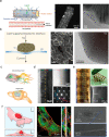
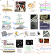
Studying the formation and interactions between biological systems and artificial materials is significant for probing complex biophysical behaviors and addressing challenging biomedical problems. Bioelectrical interfaces, especially nanostructure-based, have improved compatibility with cells and tissues and enabled new approaches to biological modulation. In particular, free-standing and remotely activated bioelectrical devices demonstrate potential for precise biophysical investigation and efficient clinical therapies. Interacting with single cells or organelles requires devices of sufficiently small size for high resolution probing. Nanoscale semiconductors, given their diverse functionalities, are promising device platforms for subcellular modulation. Tissue-level modulation requires additional consideration regarding the device's mechanical compliance for either conformal contact with the tissue surface or seamless three-dimensional (3D) biointegration. Flexible or even open-framework designs are essential in such methods. For chronic organ integration, the highest level of biocompatibility is required for both the materials and device configurations. Additionally, a scalable and high-throughput design is necessary to simultaneously interact with many individual cells in the organ. The physical, chemical, and mechanical stabilities of devices for organ implantation may be improved by ensuring matching of mechanical behavior at biointerfaces, including passivation or resistance designs to mitigate physiological impacts, or incorporating self-healing or adaptative properties. Recent research demonstrates principles of nanostructured material designs that can be used to improve biointerfaces. Nanoenabled extracellular interfaces were frequently used for either electrical or remote optical modulation of cells and tissues. In particular, methods are now available for designing and screening nanostructured silicon, especially chemical vapor deposition (CVD)-derived nanowires and two-dimensional (2D) nanostructured membranes, for biological modulation in vitro and in vivo. For intra- and intercellular biological modulation, semiconductor/cell composites have been created through the internalization of nanowires, and such cellular composites can even integrate with living tissues. This approach was demonstrated for both neuronal and cardiac modulation. At a different front, laser-derived nanocrystalline semiconductors showed electrochemical and photoelectrochemical activities, and they were used to modulate cells and organs. Recently, self-assembly of nanoscale building blocks enabled fabrication of efficient monolithic carbon-based electrodes for in vitro stimulation of cardiomyocytes, ex vivo stimulation of retinas and hearts, and in vivo stimulation of sciatic nerves. Future studies on nanoenabled bioelectrical modulation should focus on improving efficiency and stability of current and emerging technologies. New materials and devices can access new interrogation targets, such as subcellular structures, and possess more adaptable and responsive properties enabling seamless integration. Drawing inspiration from energy science and catalysis can help in such progress and open new avenues for biological modulation. The fundamental study of living bioelectronics could yield new cellular composites for diverse biological signaling control. In situ self-assembled biointerfaces are of special interest in this area as cell type targeting can be achieved.
© 2021 The Authors. Co-published by ShanghaiTech University and American Chemical Society.
Conflict of interest statement
The authors declare no competing financial interest.
Figures


Similar articles
-
Rational Design of Semiconductor Nanostructures for Functional Subcellular Interfaces.Acc Chem Res. 2018 May 15;51(5):1014-1022. doi: 10.1021/acs.accounts.7b00555. Epub 2018 Apr 18. Acc Chem Res. 2018. PMID: 29668260 Free PMC article.
-
Wearable and Implantable Soft Bioelectronics Using Two-Dimensional Materials.Acc Chem Res. 2019 Jan 15;52(1):73-81. doi: 10.1021/acs.accounts.8b00491. Epub 2018 Dec 26. Acc Chem Res. 2019. PMID: 30586292 Review.
-
Micelle-enabled self-assembly of porous and monolithic carbon membranes for bioelectronic interfaces.Nat Nanotechnol. 2021 Feb;16(2):206-213. doi: 10.1038/s41565-020-00805-z. Epub 2020 Dec 7. Nat Nanotechnol. 2021. PMID: 33288948 Free PMC article.
-
In Situ Synthesis and Assembly of Functional Materials and Devices in Living Systems.Acc Chem Res. 2024 Aug 6;57(15):2013-2026. doi: 10.1021/acs.accounts.4c00049. Epub 2024 Jul 15. Acc Chem Res. 2024. PMID: 39007720 Review.
-
Nanoelectronics-biology frontier: From nanoscopic probes for action potential recording in live cells to three-dimensional cyborg tissues.Nano Today. 2013 Aug 1;8(4):351-373. doi: 10.1016/j.nantod.2013.05.001. Nano Today. 2013. PMID: 24073014 Free PMC article.
Cited by
-
Biomaterials for neuroengineering: applications and challenges.Regen Biomater. 2025 Feb 21;12:rbae137. doi: 10.1093/rb/rbae137. eCollection 2025. Regen Biomater. 2025. PMID: 40007617 Free PMC article. Review.
-
Exploring Present and Future Directions in Nano-Enhanced Optoelectronic Neuromodulation.Acc Chem Res. 2024 May 7;57(9):1398-1410. doi: 10.1021/acs.accounts.4c00086. Epub 2024 Apr 23. Acc Chem Res. 2024. PMID: 38652467 Free PMC article. Review.
-
Nanoenabled Trainable Systems: From Biointerfaces to Biomimetics.ACS Nano. 2022 Dec 27;16(12):19651-19664. doi: 10.1021/acsnano.2c08042. Epub 2022 Dec 14. ACS Nano. 2022. PMID: 36516872 Free PMC article. Review.
-
Precise genome-editing in human diseases: mechanisms, strategies and applications.Signal Transduct Target Ther. 2024 Feb 26;9(1):47. doi: 10.1038/s41392-024-01750-2. Signal Transduct Target Ther. 2024. PMID: 38409199 Free PMC article. Review.
-
Beyond 25 years of biomedical innovation in nano-bioelectronics.Device. 2024 Jul 19;2(7):100401. doi: 10.1016/j.device.2024.100401. Device. 2024. PMID: 39119268 Free PMC article.
References
-
- Obaid A.; Hanna M. E.; Wu Y. W.; Kollo M.; Racz R.; Angle M. R.; Muller J.; Brackbill N.; Wray W.; Franke F.; Chichilnisky E. J.; Hierlemann A.; Ding J. B.; Schaefer A. T.; Melosh N. A. Massively parallel microwire arrays integrated with CMOS chips for neural recording. Sci. Adv. 2020, 6, eaay2789.10.1126/sciadv.aay2789. - DOI - PMC - PubMed
-
- Parameswaran R.; Carvalho-de-Souza J. L.; Jiang Y.; Burke M. J.; Zimmerman J. F.; Koehler K.; Phillips A. W.; Yi J.; Adams E. J.; Bezanilla F.; Tian B. Photoelectrochemical modulation of neuronal activity with free-standing coaxial silicon nanowires. Nat. Nanotechnol. 2018, 13, 260–266. 10.1038/s41565-017-0041-7. - DOI - PMC - PubMed
LinkOut - more resources
Full Text Sources
