. 2022 Mar 30;28(4):586-598.
doi: 10.1093/ibd/izab239.

Neutrophil Extracellular Trap Density Increases With Increasing Histopathological Severity of Crohn's Disease

Affiliations

Neutrophil Extracellular Trap Density Increases With Increasing Histopathological Severity of Crohn's Disease

Angie L Schroder et al. Inflamm Bowel Dis. .

Abstract

Background: Intestinal neutrophil recruitment is a characteristic feature of the earliest stages of inflammatory bowel disease (IBD). Neutrophil elastase (NE) and myeloperoxidase (MPO) mediate the formation of neutrophil extracellular traps (NETs); NETs produce the bactericidal oxidant hypochlorous acid (HOCl), causing host tissue damage when unregulated. The project aim was to investigate the relationship between NET formation and clinical IBD in humans.

Methods: Human intestinal biopsies were collected from Crohn's disease (CD) patients, endoscopically categorized as unaffected, transitional, or diseased, and assigned a histopathological score.

Results: A significant linear correlation was identified between pathological score and cell viability (TUNEL+). Immunohistochemical analysis revealed the presence of NET markers NE, MPO, and citrullinated histone (CitH3) that increased significantly with increasing histopathological score. Diseased specimens showed greater MPO+-immunostaining than control (P < .0001) and unaffected CD (P < .0001), with transitional CD specimens also showing greater staining than controls (P < .05) and unaffected CD (P < .05). Similarly, NE+-immunostaining was elevated significantly in diseased CD than controls (P < .0001) and unaffected CD (P < .0001) and was significantly higher in transitional CD than in controls (P < .0001) and unaffected CD (P < .0001). The CitH3+-immunostaining of diseased CD was significantly higher than controls (P < .05), unaffected CD (P < .0001) and transitional CD (P < .05), with transitional CD specimens showing greater staining than unaffected CD (P < .01). Multiplex immunohistochemistry with z-stacking revealed colocalization of NE, MPO, CitH3, and DAPI (cell nuclei), confirming the NET assignment.

Conclusion: These data indicate an association between increased NET formation and CD severity, potentially due to excessive MPO-mediated HOCl production in the extracellular domain, causing host tissue damage that exacerbates CD.

Keywords: inflammatory bowel disease; myeloperoxidase; neutrophil extracellular traps.

Plain language summary

Our data show for the first time that the density of neutrophil extracellular trap formed in the bowel of Crohn’s disease patients increases with increasing disease severity, suggesting that myeloperoxidase-mediated host-tissue damage may play a role in disease pathogenesis.


Conflict of interest statement

The authors have no conflicts of interest to declare.

Figures

Graphical Abstract
Graphical Abstract
Figure 1.
Figure 1.
Representative histological stained biopsy samples stained with Hematoxylin & Eosin (A) and Alcian blue (B). Images are of control tissue (C), unaffected (U), transitional (T), and diseased (D) Crohn’s disease tissue. Blue arrowhead indicates crypt dropout, green indicates edematous disruption of basement membrane, red indicates goblet cell loss. Scale bars = 100 µm (C). Pathological scoring of samples graphed as compared with the assigned grouping of each sample: <4 (healthy control, n = 3); 0–4 (unaffected, n = 5); 4.5–7 (transitional, n = 6); 7.5–10 (diseased, n = 5).
Figure 2.
Figure 2.
A, Representative images of TUNEL+ staining of frozen-sectioned healthy control and CD-affected biopsies with a fluorometric TUNEL assay. Blue: DAPI; Green: FITC—fragmented DNA. Scale bar = 100 µm. B, Linear correlation between TUNEL+ and average pixel value. Pearson correlation coefficient r2 = 0.261, P < .05.
Figure 3.
Figure 3.
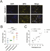
A, Representative images of neutrophil and neutrophil extracellular trap (NET) morphology observed in 7-µm sections of diseased Crohn’s patient specimens using immunofluorescence analysis of myeloperoxidase. Sample 1 represents an area of intact neutrophils in the tissue, and sample 2 represents an area where some neutrophils have undergone NET formation, indicated by white arrowheads. Scale bar = 50 µm. B, Quantification of average pixel value for n = 3-5 images collected per sample. Pixel value calculated and normalized for image number. Data categorized by assigned pathological grouping, presented as mean ± SD. Kruskal-Wallis test *P < .05, ****P < .0001. C, Linear correlation of immunofluorescence quantification data and pathological score. Pearson correlation coefficient r2 = 0.5499, P < .001.
Figure 4.
Figure 4.
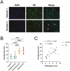
A, Representative images of neutrophil and neutrophil extracellular trap (NET) morphology observed in 7-µm sections of diseased Crohn’s patient specimens using immunofluorescence analysis of neutrophil elastase. Sample 1 represents an area of intact neutrophils in the tissue, and sample 2 represents an area where some neutrophils have undergone NET formation, indicated by white arrowheads. Scale bar = 50 µm. B, Quantification of average pixel value for n = 3-5 images collected per sample. Pixel value calculated and normalized for image number. Data categorized by assigned pathological grouping, presented as mean ± SD. Kruskal-Wallis test ****P < .0001. C, Linear correlation of immunofluorescence quantification data and pathological score. Pearson correlation coefficient r2 = 0.4495, P < .005.
Figure 5.
Figure 5.
A, Representative images of neutrophil and neutrophil extracellular trap (NET) morphology observed in 7-µm sections of diseased Crohn’s patient specimens using immunofluorescence analysis of citrullinated histone H3. Sample 1 represents an area of intact neutrophils in the tissue, and sample 2 represents an area where some neutrophils have undergone NET formation, indicated by white arrowheads. Scale bar = 50 µm. B, Quantification of average pixel value for n = 3-5 images collected per sample. Pixel value calculated and normalized for image number. Data categorized by assigned pathological grouping, presented as mean ± SD. Kruskal-Wallis test *P < .05, **P < .01 ****P < .0001. C, Linear correlation of immunofluorescence quantification data and pathological score. Pearson correlation coefficient r2 = 0.4981, P < .005.
Figure 6.
Figure 6.
Representative immunofluorescent staining of multiplex imaging of neutrophil extracellular trap (NET) structure on diseased tissue from a Crohn’s disease patient. Tissue was labeled for myeloperoxidase (yellow), neutrophil elastase (green), and citrullinated histone (red). with nuclear marker DAPI (blue). Images taken using LSM 880 confocal microscope with Airyscan processing and then images were merged with z-stacking standard software. NET structures indicated with white arrows.
Figure 7.
Figure 7.
Representative 3D image of multiplex imaging of neutrophil extracellular trap (NET) structure on diseased tissue from a Crohn’s disease patient. Tissue was labeled for myeloperoxidase (yellow), neutrophil elastase (green), and citrullinated histone (red), with nuclear marker DAPI (blue). Images taken using LSM 880 confocal microscope with Airyscan processing and then merged via z-stacking using the standard software supplied with the microscope. NET structures indicated with open arrows in the merged image while neutrophil with enzymes translocated to nucleus indicated with solid (black) arrow.

Comment in

References

    1. Chernavskaia O, Heuke S, Vieth M, et al. Beyond endoscopic assessment in inflammatory bowel disease: real-time histology of disease activity by non-linear multimodal imaging. Sci Rep. 2016;6:29239. - PMC - PubMed
    1. Chami B, Martin NJJ, Dennis JM, Witting PK. Myeloperoxidase in the inflamed colon: A novel target for treating inflammatory bowel disease. Arch Biochem Biophys. 2018;645:61–71. - PubMed
    1. Magro F, Langner C, Driessen A, et al. ; European Society of Pathology (ESP); European Crohn’s and Colitis Organisation (ECCO). . European consensus on the histopathology of inflammatory bowel disease. J Crohn’s Colitis. 2013;7:827–851. - PubMed
    1. Kim W-H. Atlas of Inflammatory Bowel Diseases. Berlin, Heidelberg: Springer Berlin Heidelberg; 2015.
    1. Fournier BM, Parkos CA. The role of neutrophils during intestinal inflammation. Mucosal Immunol. 2012;5:354–366. - PubMed

Publication types