. 2021 Dec;78(23):7873-7898.
doi: 10.1007/s00018-021-03993-6. Epub 2021 Nov 1.

Role of Akt isoforms in neuronal insulin signaling and resistance

Affiliations

Role of Akt isoforms in neuronal insulin signaling and resistance

Medha Sharma et al. Cell Mol Life Sci. 2021 Dec.

Abstract

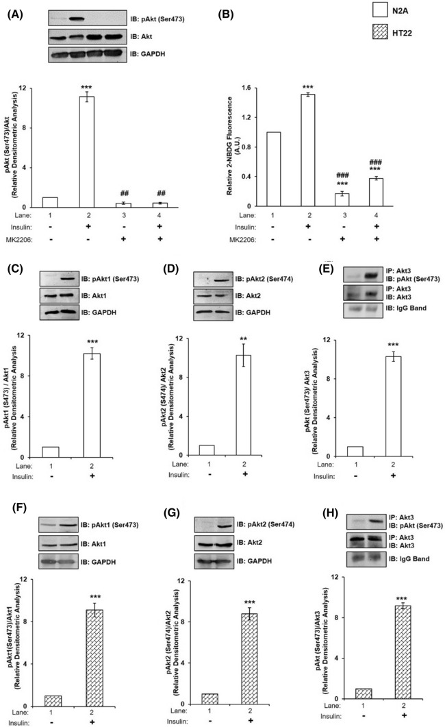
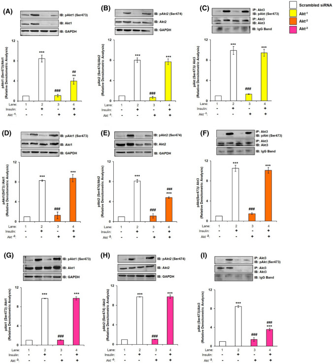
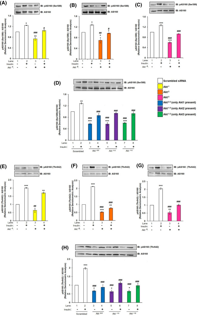
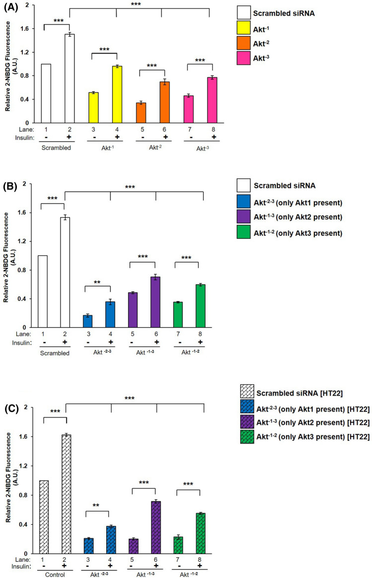
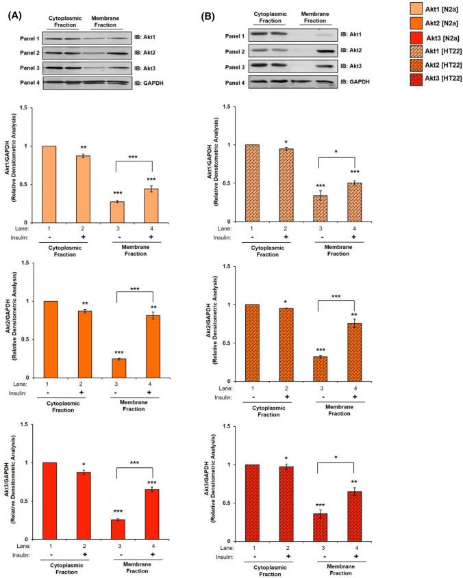
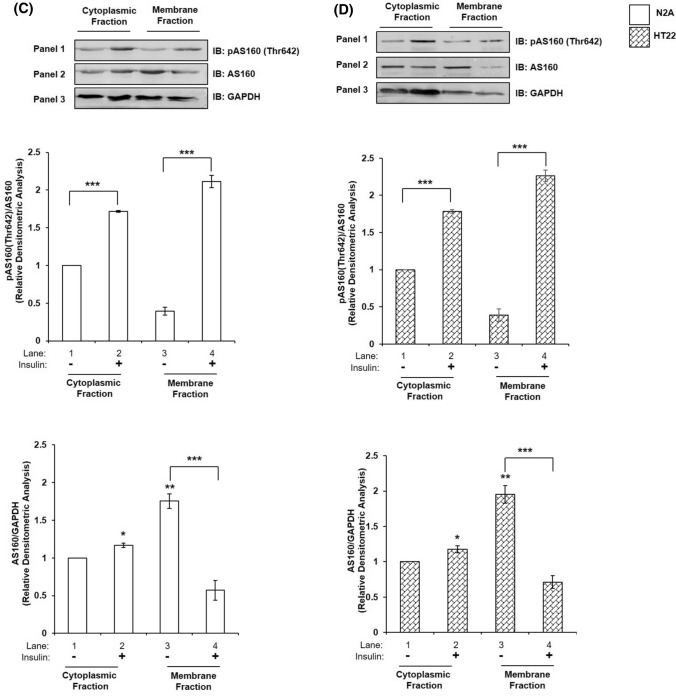
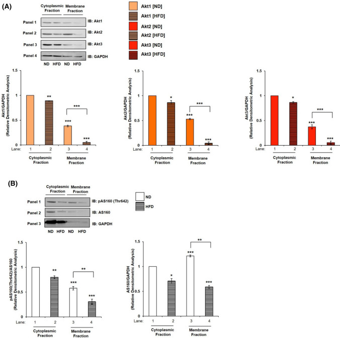
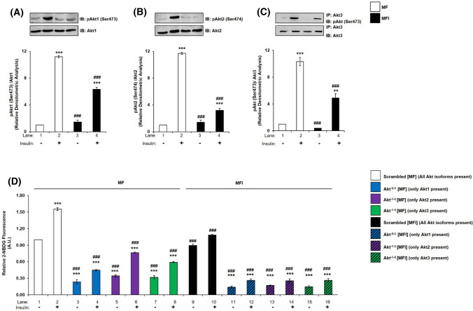
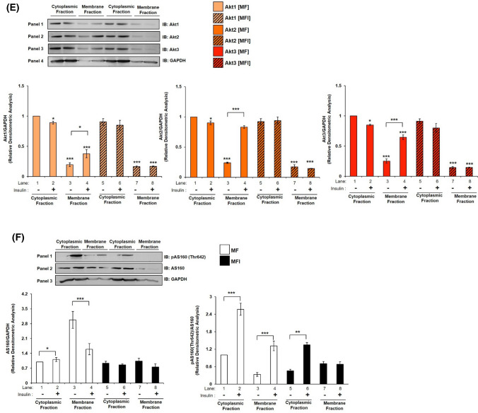
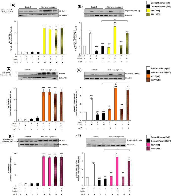
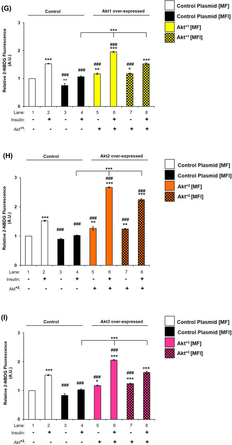
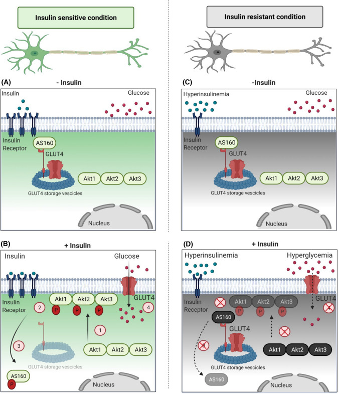
The aim of the present study was to determine the role of Akt isoforms in insulin signaling and resistance in neuronal cells. By silencing Akt isoforms individually and in pairs, in Neuro-2a and HT22 cells we observed that, in insulin-sensitive condition, Akt isoforms differentially reduced activation of AS160 and glucose uptake with Akt2 playing the major role. Under insulin-resistant condition, phosphorylation of all isoforms and glucose uptake were severely affected. Over-expression of individual isoforms in insulin-sensitive and resistant cells differentially reversed AS160 phosphorylation with concomitant reversal in glucose uptake indicating a compensatory role of Akt isoforms in controlling neuronal insulin signaling. Post-insulin stimulation Akt2 translocated to the membrane the most followed by Akt3 and Akt1, decreasing glucose uptake in the similar order in insulin-sensitive cells. None of the Akt isoforms translocated in insulin-resistant cells or high-fat-diet mediated diabetic mice brain cells. Based on our data, insulin-dependent differential translocation of Akt isoforms to the plasma membrane turns out to be the key factor in determining Akt isoform specificity. Thus, isoforms play parallel with predominant role by Akt2, and compensatory yet novel role by Akt1 and Akt3 to regulate neuronal insulin signaling, glucose uptake, and insulin-resistance.

Keywords: AS160; Akt1; Akt2; Akt3; Neuronal insulin resistance; Neuronal insulin signaling.


Conflict of interest statement

The authors declare no conflicts of interest.

Figures

Fig. 1
Fig. 1
Effect of Akt inhibition on neuronal insulin signaling and glucose uptake; and effect of insulin on Akt and its isoforms. A N2A cells were differentiated in 2% DMSO for 3 days and stimulated with or without 30 µM MK2206 and/or 100 nM insulin for 30 min as indicated. Cells were lysed and subjected to western blotting, followed by probing with relevant primary antibodies. Bar represents relative change in pAkt (Ser-473) probed with anti-Akt antibody. B Differentiated N2A cells were serum starved for 2 h, followed by 100 nM insulin for 30 min. Uptake of 2-NBDG was then measured. Bar represents relative change in uptake of 2-NBDG. CE N2A cells were differentiated in 2% DMSO for 3 days and stimulated with or without 100 nM insulin for 30 min. Cells were lysed and subjected to western blotting, followed by probing with relevant primary antibodies. C Bar represents relative change in pAkt1 (Ser-473) probed with anti-Akt1 antibody. D Bar represents relative change in pAkt2 (Ser-474) probed with anti-Akt2 antibody. E Post-insulin stimulation, lysates were subjected to immunoprecipitation using anti-Akt3 antibody. Bar represents relative change in pAkt (Ser-473) probed with anti-Akt3 antibody. FH HT22 cells were differentiated in neurobasal media containing 2 mmol/L glutamine and 1 × N2 supplement for 48 h and stimulated with or without 100 nM insulin for 30 min. Cells were lysed and subjected to western blotting, followed by probing with relevant primary antibodies. F Bar represents relative change in pAkt1 (Ser-473) probed with anti-Akt1 antibody. G Bar represents relative change in pAkt2 (Ser-474) probed with anti-Akt2 antibody. H Post-insulin stimulation, lysates were subjected to immunoprecipitation using anti-Akt3 antibody. Bar represents relative change in pAkt (Ser-473) probed with anti-Akt3 antibody. GAPDH has been used as a loading control (A, C, D, F, G). IgG band was used as loading control (E, H). Experiments were executed three times and a representative result is shown. Data expressed are mean ± SE. ***P < 0.001, **P < 0.01 compared to lane 1, ###P < 0.001, ##P < 0.01 compared to lane 2. IP Immunoprecipitation; IB Immunoblot; A.U Arbitrary Units
Fig. 2
Fig. 2
Effect of silencing of one isoform, on expression and phosphorylation of other isoform in N2A cells. Three days post-proliferation, N2A cells were transfected with Akt1, Akt2 or Akt3 specific siRNA and then differentiated in 2% DMSO for 3 days. Cells were stimulated with or without 100 nM insulin for 30 min prior to cell lysis. Lysate was subjected to western blotting, followed by probing with relevant primary antibodies. A, D, G Bar represents relative change in pAkt1 (Ser-473) when probed with anti-Akt1 antibody. B, E, H Bar represents relative change in pAkt2 (Ser-474) when probed with anti-Akt2 antibody. C, F, I Bar represents relative change in pAkt (Ser-473) when probed with anti-Akt3 antibody. GAPDH has been used as a loading control (A, B, D, E, G, H). IgG band was used as loading control (C, F, I). Experiments were executed three times and a representative result is shown. Data expressed are mean ± SE. ***P < 0.001, **P < 0.01 as compared to lane 1; ###P < 0.001, ##P < 0.01 as compared to lane 2. IP Immunoprecipitation; IB Immunoblot
Fig. 3
Fig. 3
Effect of Akt isoform silencing on AS160 in N2A cells. Three days post-proliferation, N2A cells were transfected with single (Akt−1, Akt−2, and Akt−3) or double (Akt−2–3, Akt−1–3 or Akt−1–2) specific siRNA and then differentiated in 2% DMSO for 3 days. Cells were stimulated with or without 100 nM insulin for 30 min prior to cell lysis. Lysate was subjected to western blotting, followed by probing with relevant primary antibodies. (AD) Bar represents relative change in pAS160 (Ser-588) when probed with anti-AS160 antibody. (EH) Bar represents relative change in pAS160 (Thr-642) when probed with anti-AS160 antibody. Experiments were executed three times and a representative result is shown. Data expressed are mean ± SE. ***P < 0.001, **P < 0.01, *P < 0.05 as compared to lane 1; ###P < 0.001, ##P < 0.01, #P < 0.05 as compared to lane 2. IB Immunoblot
Fig. 4
Fig. 4
Effect of Akt isoform silencing on glucose uptake. Three days post-proliferation, cells were transfected with (A) Single (Akt−1, Akt−2 or Akt−3) (N2A) (B) Double (Akt−2–3, Akt−1–3, Akt−1–2) (N2A) (C) Double (Akt−2–3, Akt−1–3, Akt−1–2) (HT22) specific siRNA and then differentiated for 3 days. Differentiated cells were serum starved for 2 h, followed by 100 nM insulin for 30 min. Uptake of 2-NBDG was then measured. Bar represents relative change in uptake of 2-NBDG. Experiments were executed three times and a representative result is shown. Data expressed are mean ± SE. ***P < 0.001, **P < 0.01 as indicated
Fig. 5
Fig. 5
Mechanism underlying Akt isoform specificity in neuronal cells. A N2A cells were differentiated in 2% DMSO for 3 days. Three days post-differentiation, membrane and cytosol fraction were isolated and subjected to western blotting, followed by probing with relevant primary antibodies. Bar represents relative change in Akt1, Akt2 or Akt3 when probed with anti-Akt isoform specific antibody. B HT22 cells were differentiated in neurobasal media containing 2 mmol/L glutamine and 1 × N2 supplement for 48 h. Three days post-differentiation, membrane and cytosol fraction were isolated and subjected to western blotting, followed by probing with relevant primary antibodies. Bar represents relative change in Akt1, Akt2 or Akt3 when probed with anti-Akt isoform specific antibody. C, D Bar represents relative change in AS160 when probed with anti-AS160 antibody (N2A/HT22 as indicated). Bar represents relative change in pAS160 (Thr-642) when probed with anti-AS160 antibody (N2A/HT22 as indicated). GAPDH has been used as a loading control. Experiments were executed three times and a representative result is shown. Data expressed are mean ± SE. ***P < 0.001, **P < 0.01, *P < 0.05 compared to lane 1 or as indicated. IB Immunoblot
Fig. 5
Fig. 5
Mechanism underlying Akt isoform specificity in neuronal cells. A N2A cells were differentiated in 2% DMSO for 3 days. Three days post-differentiation, membrane and cytosol fraction were isolated and subjected to western blotting, followed by probing with relevant primary antibodies. Bar represents relative change in Akt1, Akt2 or Akt3 when probed with anti-Akt isoform specific antibody. B HT22 cells were differentiated in neurobasal media containing 2 mmol/L glutamine and 1 × N2 supplement for 48 h. Three days post-differentiation, membrane and cytosol fraction were isolated and subjected to western blotting, followed by probing with relevant primary antibodies. Bar represents relative change in Akt1, Akt2 or Akt3 when probed with anti-Akt isoform specific antibody. C, D Bar represents relative change in AS160 when probed with anti-AS160 antibody (N2A/HT22 as indicated). Bar represents relative change in pAS160 (Thr-642) when probed with anti-AS160 antibody (N2A/HT22 as indicated). GAPDH has been used as a loading control. Experiments were executed three times and a representative result is shown. Data expressed are mean ± SE. ***P < 0.001, **P < 0.01, *P < 0.05 compared to lane 1 or as indicated. IB Immunoblot
Fig. 6
Fig. 6
Effect of insulin on subcellular translocation of GLUT4 and AS160. I Three days post-proliferation, N2A cells differentiated in 2% DMSO for 3 days. Cells were stimulated with or without 100 nM insulin for 30 min followed by fixation and permeabilization and probed with anti-GLUT4 and anti-AS160 antibody. Cells were subjected to immunofluorescence microscopy using anti-goat Alexa 555 and anti-rabbit CFL 488 secondary antibodies, respectively. Bar corresponds to 10 μm. Images were captured from different fields and a representative image of 3 images is presented. Red arrow indicates GLUT4, green arrow indicates AS160 and yellow arrow indicates co-localized GLUT4 and AS160. II Graphical representation of co-localization by Pearson’s coefficient (*P < 0.05)
Fig. 7
Fig. 7
Mechanism underlying Akt isoforms specificity in mice whole brain tissue. A Subcellular translocation of Akt isoforms in mice whole brain tissue. Mice whole brain was lysed, membrane and cytosol fraction were isolated and subjected to western blotting, followed by probing with relevant primary antibodies. Bar represents relative change in Akt1, Akt2 or Akt3 when probed with anti-Akt isoform specific antibody. Experiments were executed with three independent animals and a representative result is shown. B Subcellular redistribution of AS160 post-insulin stimulation. Bar represents relative change in AS160 when probed with anti-AS160 antibody. Bar represents relative change in pAS160 (Thr-642) when probed with anti-AS160 antibody. GAPDH has been used as a loading control. Experiments were executed with three independent animals and a representative result is shown. Data expressed are mean ± SE. ***P < 0.001, **P < 0.01, *P < 0.05 compared to lane 1 or as indicated. (ND Normal Diet; HFD High-Fat-Diet). IB Immunoblot
Fig. 8
Fig. 8
Expression and activation of Akt1, Akt2, and Akt3, glucose uptake and subcellular translocation under insulin-resistant condition in neuronal cells. (AC) N2A cells were differentiated in serum-free medium in the absence of (MF) or chronic presence of 100 nM insulin (MFI) for 3 days. Cells were lysed and subjected to western blotting, followed by probing with relevant primary antibodies. Bar represents relative change when probed with anti-Akt isoform specific antibody. A Bar represents relative change in pAkt1 (Ser-473) probed with anti-Akt1 antibody. B Bar represents relative change in pAkt2 (Ser-474) probed with anti-Akt2 antibody. C Post-insulin stimulation, lysates were subjected to immunoprecipitation using anti-Akt3 antibody. Bar represents relative change in pAkt (Ser-473) probed with anti-Akt3 antibody. D Three days post-proliferation, N2A cells were transfected with double (Akt−2–3, Akt−1–3 or Akt−1–2) specific siRNA and then differentiated under MF MFI condition for 3 days. Differentiated N2A cells were serum starved for 2 h, followed by 100 nM insulin for 30 min. Uptake of 2-NBDG was then measured. E Subcellular translocation of Akt isoforms post-insulin stimulation. N2A cells were differentiated under MF MFI condition for 3 days and stimulated with or without 100 nM insulin for 30 min as indicated. Cells were lysed, and membrane and cytosol fraction were isolated and subjected to western blotting, followed by probing with relevant primary antibodies. Bar represents relative change in Akt1, Akt2 or Akt3 when probed with anti-Akt isoform specific antibody. F Subcellular translocation of AS160 post-insulin stimulation. Bar represents relative change in pAS160 (Thr-642) when probed with anti-AS160 antibody. GAPDH has been used as a loading control. Experiments were executed three times and a representative result is shown. Data expressed are mean ± SE. ***P < 0.001, **P < 0.01, *P < 0.05 compared to lane 1 or as indicated; ###P < 0.001, ##P < 0.01, #P < 0.05 compared to lane 2. IP Immunoprecipitation; IB Immunoblot; A.U Arbitrary Units
Fig. 8
Fig. 8
Expression and activation of Akt1, Akt2, and Akt3, glucose uptake and subcellular translocation under insulin-resistant condition in neuronal cells. (AC) N2A cells were differentiated in serum-free medium in the absence of (MF) or chronic presence of 100 nM insulin (MFI) for 3 days. Cells were lysed and subjected to western blotting, followed by probing with relevant primary antibodies. Bar represents relative change when probed with anti-Akt isoform specific antibody. A Bar represents relative change in pAkt1 (Ser-473) probed with anti-Akt1 antibody. B Bar represents relative change in pAkt2 (Ser-474) probed with anti-Akt2 antibody. C Post-insulin stimulation, lysates were subjected to immunoprecipitation using anti-Akt3 antibody. Bar represents relative change in pAkt (Ser-473) probed with anti-Akt3 antibody. D Three days post-proliferation, N2A cells were transfected with double (Akt−2–3, Akt−1–3 or Akt−1–2) specific siRNA and then differentiated under MF MFI condition for 3 days. Differentiated N2A cells were serum starved for 2 h, followed by 100 nM insulin for 30 min. Uptake of 2-NBDG was then measured. E Subcellular translocation of Akt isoforms post-insulin stimulation. N2A cells were differentiated under MF MFI condition for 3 days and stimulated with or without 100 nM insulin for 30 min as indicated. Cells were lysed, and membrane and cytosol fraction were isolated and subjected to western blotting, followed by probing with relevant primary antibodies. Bar represents relative change in Akt1, Akt2 or Akt3 when probed with anti-Akt isoform specific antibody. F Subcellular translocation of AS160 post-insulin stimulation. Bar represents relative change in pAS160 (Thr-642) when probed with anti-AS160 antibody. GAPDH has been used as a loading control. Experiments were executed three times and a representative result is shown. Data expressed are mean ± SE. ***P < 0.001, **P < 0.01, *P < 0.05 compared to lane 1 or as indicated; ###P < 0.001, ##P < 0.01, #P < 0.05 compared to lane 2. IP Immunoprecipitation; IB Immunoblot; A.U Arbitrary Units
Fig. 9
Fig. 9
Effect of Akt1, Akt2, and Akt3 over-expression on expression and activation of AS160, and neuronal glucose uptake under insulin-resistant condition in neuronal cells. AF Three days post-proliferation, Akt1, Akt2 or Akt3 were over-expressed using isoform specific plasmids. N2A cells were differentiated in serum-free medium in the absence of (MF) or chronic presence of 100 nM insulin (MFI) for 3 days. Cells were lysed and subjected to western blotting, followed by probing with relevant primary antibodies. Bar represents relative change when probed with anti-Akt isoform specific antibody. A, C, E Bar represents relative change in Akt1/Akt2/Akt3 probed with anti-Akt isoform specific antibody. GAPDH was used as loading control B, D, F Bar represents relative change in pAS160 (Thr-642) when probed with anti-AS160 antibody. GI Three days post-proliferation, Akt1, Akt2 or Akt3 were over-expressed using isoform specific plasmids. N2A cells were differentiated in serum-free medium in the absence of (MF) or chronic presence of 100 nM insulin (MFI) for 3 days. N2A cells were serum starved for 2 h, followed by 100 nM insulin for 30 min. Uptake of 2-NBDG was then measured. Experiments were executed three times and a representative result is shown. Data expressed are mean ± SE. ***P < 0.001, **P < 0.01, *P < 0.05 compared to lane 1 or as indicated, ###P < 0.001, ##P < 0.01, #P < 0.05 compared to lane 2. IB Immunoblot; A.U Arbitrary Units
Fig. 9
Fig. 9
Effect of Akt1, Akt2, and Akt3 over-expression on expression and activation of AS160, and neuronal glucose uptake under insulin-resistant condition in neuronal cells. AF Three days post-proliferation, Akt1, Akt2 or Akt3 were over-expressed using isoform specific plasmids. N2A cells were differentiated in serum-free medium in the absence of (MF) or chronic presence of 100 nM insulin (MFI) for 3 days. Cells were lysed and subjected to western blotting, followed by probing with relevant primary antibodies. Bar represents relative change when probed with anti-Akt isoform specific antibody. A, C, E Bar represents relative change in Akt1/Akt2/Akt3 probed with anti-Akt isoform specific antibody. GAPDH was used as loading control B, D, F Bar represents relative change in pAS160 (Thr-642) when probed with anti-AS160 antibody. GI Three days post-proliferation, Akt1, Akt2 or Akt3 were over-expressed using isoform specific plasmids. N2A cells were differentiated in serum-free medium in the absence of (MF) or chronic presence of 100 nM insulin (MFI) for 3 days. N2A cells were serum starved for 2 h, followed by 100 nM insulin for 30 min. Uptake of 2-NBDG was then measured. Experiments were executed three times and a representative result is shown. Data expressed are mean ± SE. ***P < 0.001, **P < 0.01, *P < 0.05 compared to lane 1 or as indicated, ###P < 0.001, ##P < 0.01, #P < 0.05 compared to lane 2. IB Immunoblot; A.U Arbitrary Units
Fig. 10
Fig. 10
Schematic diagram depicting role of Akt isoforms in regulating neuronal insulin signaling. A In an insulin-sensitive, under unstimulated condition, Akt (Akt1, Akt2, and Akt3) are present in the cytoplasm. AS160 binds to GSVs (GLUT4 Storage Vesicles) and tethers GLUT4. This does not allow GLUT4 exocytosis under basal conditions. B In an insulin-sensitive, insulin stimulated condition, Akt translocates to plasma membrane in an isoform specific insulin-dependent way (1), getting phosphorylated there (2). In neuronal system, all Akt isoforms translocate in the order Akt2 > Akt3 > Akt1. An activated Akt phosphorylates AS160, hence inactivating it. Phosphorylated and thus inactivated AS160 translocated to cytoplasm (3), promoting GLUT4 dissociation from GSVs and allowing glucose uptake (4). C In an insulin-resistant, unstimulated condition, hyperinsulinemia occurs due to defects in insulin signaling. D In an insulin-resistant, insulin stimulated condition, hyperinsulinemia triggers insulin receptor down-regulation, not allowing Akt isoform specific translocation to the plasma membrane (1). This leads to inadequate phosphorylation (2), leading to Akt’s inability to phosphorylate AS160. Unphosphorylated, thus, active AS160 continues tethering GLUT4 to GSVs, not allowing GLUT4 exocytosis, hence affecting neuronal glucose uptake. (Created with BioRender.com)

References

    1. Gonzalez E, McGraw TE. The Akt kinases: isoform specificity in metabolism and cancer. Cell Cycle. 2009;8:2502–2508. - PMC - PubMed
    1. Hay N. Akt isoforms and glucose homeostasis—the leptin connection. Trends Endocrinol Metab. 2011;22:66–73. - PMC - PubMed
    1. Manning BD, Toker A. AKT/PKB signaling: navigating the network. Cell. 2017;169:381–405. - PMC - PubMed
    1. Huang X, Liu G, Guo J, Su Z. The PI3K/AKT pathway in obesity and type 2 diabetes. Int J Biol Sci. 2018;14:1483–1496. - PMC - PubMed
    1. Gupta A, Bisht B, Dey CS. Focal adhesion kinase negatively regulates neuronal insulin resistance. Biochim Biophys Acta. 2012;1822:1030–1037. - PubMed

MeSH terms