Review
. 2021 Nov 1;35(21-22):1403-1430.
doi: 10.1101/gad.348897.121.

Sentinels of chromatin: chromodomain helicase DNA-binding proteins in development and disease

Affiliations
Review

Sentinels of chromatin: chromodomain helicase DNA-binding proteins in development and disease

Andrej Alendar et al. Genes Dev. .

Abstract

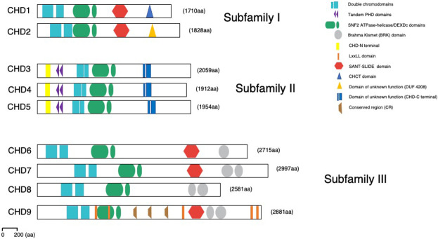
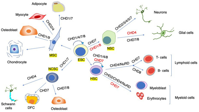
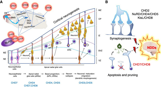
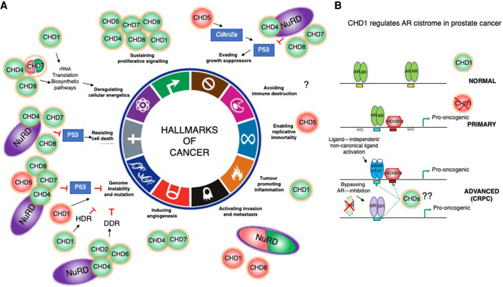
Chromatin is highly dynamic, undergoing continuous global changes in its structure and type of histone and DNA modifications governed by processes such as transcription, repair, replication, and recombination. Members of the chromodomain helicase DNA-binding (CHD) family of enzymes are ATP-dependent chromatin remodelers that are intimately involved in the regulation of chromatin dynamics, altering nucleosomal structure and DNA accessibility. Genetic studies in yeast, fruit flies, zebrafish, and mice underscore essential roles of CHD enzymes in regulating cellular fate and identity, as well as proper embryonic development. With the advent of next-generation sequencing, evidence is emerging that these enzymes are subjected to frequent DNA copy number alterations or mutations and show aberrant expression in malignancies and other human diseases. As such, they might prove to be valuable biomarkers or targets for therapeutic intervention.

Keywords: cancer; chromatin remodeling; chromodomain helicase DNA-binding proteins; development and disease.


Figures

Figure 1.
Figure 1.
Structural domain organization of the CHD enzymes. Domain representation of all three CHD subfamilies as indicated in the text. All CHDs have double chromodomains (blue squares) and SNF2-like ATPase/DEXDc helicase domains (green ovals). Subfamily II members contain tandem PHD Zn finger-like domains (purple triangles). Subfamily III members have additional Brahma Kismet (BRK) domains at the C terminus (gray oval); enzymes of both subfamilies I and III contain SANT-SLIDE domains at C terminus (red hexagon). These domains facilitate engagement and stabilization of the interaction with the chromatin substrate and allow efficient ATP hydrolysis and DNA translocation. Recently, a number of novel structural domains have been identified: CHD-N-terminal, CHD-C-terminal, CHCT, DUF4208, and conserved region (CR). CHD9 associates with nuclear receptors through LxxLL recognition motifs (orange stripes).
Figure 2.
Figure 2.
Autoinhibitory regulation and nucleosome sliding mechanism of SF2-type chromatin remodelers. (A) The ATPase motor of CHD enzymes is comprised of two RecA-like lobes ([red] lobe1, [blue] lobe2) that adopt a closed confirmation to form a contiguous surface that allows nucleic acid binding and efficient ATP hydrolysis. Biophysical and structural studies of yChd1, CHD2, and CHD4 revealed autoinhibitory domain organization whereby flanking domains fold onto the ATPase motor to prevent activation by improper substrates (Hauk et al. 2010; Ryan et al. 2011; Morra et al. 2012; Watson et al. 2012; Liu et al. 2014a). The N-terminal tandem chromodomains (CD; green) of yChd1 contain a linker helix with several conserved acidic residues that stacks onto the DNA-binding surface of lobe2 and negatively regulates yChd1 activity (Hauk and Bowman 2011; Narlikar et al. 2013). Full inhibition is achieved through folding of the “bridge” inhibitory domain adjacent to the ATPase motor (Bridge; orange). Moreover, autoinhibitory interaction of the ATPase motor with the C-terminal domains uncouples ATP hydrolysis from productive DNA translocation of Chd1p and CHD4, respectively (Hauk et al. 2010; Morra et al. 2012; Watson et al. 2012). Upon chromatin binding, interactions of CHD domains with linker DNA and protruding histone H4 tails (H4; gray) trigger major conformational change and dislodging of the autoinhibitory domains, allowing the ATPase motor to adopt an active conformation (Hauk and Bowman 2011; Nodelman et al. 2021). The interplay between the ATPase motor and flanking domains not only provides means for autoinhibition but also can promote substrate recognition and ATPase activity. In the case of CHD4, the N-terminal tandem chromodomains and PHD domains stimulate ATPase motor activity and DNA translocation (Morra et al. 2012; Watson et al. 2012), whereas the respective C-terminal DNA-binding regions of CHD1, CHD2, and CHD7 stimulate activity of the ATPase motor (Ryan and Owen-Hughes 2011; Bouazoune and Kingston 2012; Liu et al. 2014a). Thus, intradomain allosteric regulation keeps the enzyme in an inactive conformation to ensure substrate specificity, while on the other hand facilitating ATPase motor activity and DNA translocation. (GATED) yChd1 closed confirmation, (UNGATED) open confirmation, (light blue) nucleosome core, (gray) wrapped DNA. Features required for the efficient ATP binding and hydrolysis in lobe1 and lobe2 are indicated: ATP binding (magenta circles), ATP hydrolysis (green lines), and nucleic acid binding surfaces on each lobe (opaque areas) (Hauk and Bowman 2011). Top panel cartoon adapted with permission from Springer Nature from Mueller-Planitz et al. (2013), © 2013. Bottom panel republished with permission of Elsevier Science and Technology Journals from Hauk and Bowman (2011); permission conveyed through Copyright Clearance Center, Inc. (B) Historically, nucleosome crystal structure and earlier models of nucleosome sliding revealed that nucleosomal DNA can accommodate an extra base pair at the internal DNA location termed superhelix location 2 (SHL2) (Luger et al. 1997), which is a docking site for many ATP-dependent chromatin remodelers. Recent evidence shows that CHD, ISWI, and Snf2-type remodelers shift DNA discontinuously around the nucleosome (Farnung et al. 2017, 2020; Winger et al. 2018; Sabantsev et al. 2019; Yan and Chen 2020; Zhong et al. 2020; Nodelman et al. 2021). Remarkably, in an open conformation, the ATPase motor bound at the nucleosomal SHL2 position pulls 1–2 bp of DNA at the nucleosome entry site, creating a shift of only one DNA strand (tracking strand) with respect to the other (guide strand), resulting in a twist defect. Subsequent ATP binding induces closed confirmation of the ATPase motor, pulling the entire base pair of the DNA at SHL2 and forcing the DNA twist defect toward the nucleosomal exit site. Upon ATP hydrolysis, the motor readopts the open confirmation again, priming the nucleosome for the next translocation cycle. This sequential, ATP-driven conformational transition between open and closed states propels the DNA around the nucleosome histone core (Liu et al. 2017; Farnung et al. 2017, 2020; Armache et al. 2019; Nodelman et al. 2021; Yan and Chen 2020). Republished with permission of Elsevier Science and Technology Journals from Bowman (2019); permission conveyed through Copyright Clearance Center, Inc.
Figure 3.
Figure 3.
Regulation of the transcription cycle. (A) CHD enzymes are involved at different stages of transcription: initiation, promoter escape, elongation, splicing, and termination, as well as higher-order chromatin organization. The promoter nucleosome-free region (NFR) is demarcated by the ±1 nucleosomes (purple). CHDs also regulate nucleosome accessibility at enhancer elements and modulate CTCF-mediated chromatin organization. (TSS) Transcription start site, (TTS) transcription termination site, (TF) transcription factors, (GTFs) general transcription factors, (U1/2/4/5/6) spliceosome, (orange rectangle) enhancer elements, (blue rectangle) CTCF-binding site. RNA polymerase II (RNAPII) phosphorylation status of the C-terminal domain is represented by the red (serine 5 phosphorylation-paused RNAPII) and green (serine 2 phosphorylation-elongating RNAPII) circles. (B) CHD enzymes can both promote and inhibit transcription of different subsets of genes in a context-dependent manner. CHD8 and linker histone H1 (blue circle) form a trimeric repressive complex with either P53 or β-catenin (green oval) to repress target gene expression.
Figure 4.
Figure 4.
Cell fate specification and lineage commitment. CHD functions are important in diverse stem cell populations (annotated by yellow rectangles). Some CHDs are important for the emergence and self-renewal of stem cells (circular arrow), and others are vital for lineage commitment and differentiation (straight arrows). Distinct CHDs can promote (black) or inhibit (red) differentiation. For simplicity, not all intermediate precursors and terminally differentiated cells are depicted. (ESC) Embryonic stem cell, (HSC) hematopoietic stem cell, (MSC) mesenchymal stem cell, (NSC) neural stem cell, (NCSC) neural crest stem cell, (DFC) dental follicle cells.
Figure 5.
Figure 5.
CHDs regulate different aspects of neurodevelopment. (A, top) CHD enzymes are vital for the development and functionality of different parts of the brain: the cerebral neocortex (CHD2, CHD3/4/5/NuRD, CHD7, and CHD8), cerebellum (CHD4/NuRD, CHD6, CHD7, and CHD8), hypothalamus (CHD2), thalamus (CHD8), and hippocampus (CHD4/5/NuRD, CHD7, and CHD8). They regulate neural stem cell niches in the cerebrum (subventricular zone [SVZ]) and hippocampus (subgranular zone of the dentate gyrus [SGZ/DG]) and are essential for proper neuronal differentiation and migration. CHD7 is important for the development of the inner ear and olfactory system and regulates migration of the neuroblasts that emerge from the neural stem cells in the SVZ and proceed through the rostral migratory stream (RMS) to the olfactory bulb (OB). The cartoon represents the sagittal section of the rodent brain and highlights CHD functions in the respective brain regions (lines). (Bottom) CHDs are required at various stages of cortical neurogenesis. During the early stages of cortical neurogenesis, symmetrical division of the neuroepithelial cells (NEs) in the ventricular zone (VZ) expands their pool, whereas asymmetrical division gives rise to apical neural precursor cells (NPCs), such as apical radial glial cells (aRGs), and pioneer neurons. Apical progenitor cells give rise to neurons through basal intermediate precursors (bIP) and radial glial cells (bRG) that reside in the subventricular zone (SVZ). Neurons use aRG and bRG fibers to migrate to the specified layer in the cortical plate (CP), where they establish synaptic connections with subplate layer neurons during maturation. CHD remodelers involved at distinct stages are highlighted bellow. (MZ) Marginal zone, (IZ) intermediate zone. Cortical neurogenesis cartoon representation adapted with permission from Sokpor et al. (2018). (B, top) Synaptogenesis between neurons initiates during embryogenesis, but synaptic plasticity proceeds throughout life. Synaptic plasticity is the property of synapses to strengthen or weaken in response to changes in both the amplitude and the temporal dynamics of neuronal activity. Sensory inputs and intrinsic brain activity can effect long-term changes in synaptic efficacy and eventually increase or decrease neuronal connectivity by modulating the number of synapses (Bourgeron 2015). Some CHD enzymes regulate neuronal connectivity (CHD4, CHD5, and CHD8) and excitability (CHD2 and CHD8), whereas others regulate synaptic vesicle recycling (KisL) and expression of genes essential for synaptic homeostasis and plasticity (CHD8). (Bottom) Apoptosis and pruning are required for the maintenance and refinement of the neural circuitries, and CHD7/8 inhibition of P53-mediated apoptosis plays an active role in this process. Dysregulation of CHD enzyme function affects synaptogenesis and apoptosis and leads to aberrant neurogenesis and connectivity, which results in the emergence of diverse neurodevelopmental disorders (NDDs).
Figure 6.
Figure 6.
The role of CHDs in tumorigenesis. (A) CHD enzymes are context-dependent tumor suppressors (TS; red) and oncogenes (green) that contribute to acquisition of different hallmarks of cancer. They can drive tumorigenesis by directly promoting transcription of oncogenes and repression of TS genes (CDKN2A and TP53; blue rectangles). Some enzymes can exert both oncogenic and TS function (CHD1, CHD4/NuRD, CHD7, and CHD8). For simplicity, not all players that are mentioned in the text are indicated in the cartoon. CHD functions in human malignancies are still largely unexplored, and it is likely that all members are dysregulated and mutated in diverse cancers and contribute to different aspects of tumorigenesis. Cartoon representation adapted from Hanahan and Weinberg (2011) with permission from Elsevier. (B, top) CHD1 nucleosome remodeling activity restricts binding of AR (green ovals) to prostetic lineage enhancers (yellow rectangles) in normal prostates. (Middle) During prostate tumorigenesis, in primary tumors, CHD1 loss leads to AR cistrome redistribution to a different subset of enhancers (blue rectangles), with HOXB13 (red hexagon) binding motifs (red squares) to drive expression of pro-oncogenic gene networks. (Bottom) More advanced, castration-resistant prostate cancers (CRPCs) are characterized by amplifications or mutations in the AR ligand-binding domain (LBD; AR-V7 as an example; blue ovals) (Jeselsohn et al. 2015; Watson et al. 2015). The LBD mutations allow promiscuous activation by noncanonical ligands (adrenal androgens, estrogen, progesterone, and glucocorticoids) to secure sustained expression of a subset of critical AR target genes (Watson et al. 2015). Additionally, antiandrogen-resistant prostate cancers can fully bypass the requirement for AR signaling by activating GR-mediated (violet ovals) transcriptional regulation of the AR cistrome (Arora et al. 2013). The role of other CHDs in the regulation of alternative transcriptional pathways that sustain expression of the AR-driven oncogenic gene networks in CRPC prostate cancers is unknown.

References

    1. Akhtar A, Zink D, Becker PB. 2000. Chromodomains are protein-RNA interaction modules. Nature 407: 405–409. 10.1038/35030169 - DOI - PubMed
    1. Alendar A, Lambooij J-P, Bhaskaran R, Lancini C, Song J-Y, Vugt Hv, Snoek M, Berns A. 2020. Gene expression regulation by the chromodomain helicase DNA-binding protein 9 (CHD9) chromatin remodeler is dispensable for murine development. PLoS One 15: e0233394. 10.1371/journal.pone.0233394 - DOI - PMC - PubMed
    1. Allen MD, Religa TL, Freund SMV, Bycroft M. 2007. Solution structure of the BRK domains from CHD7. J Mol Biol 371: 1135–1140. 10.1016/j.jmb.2007.06.007 - DOI - PubMed
    1. Arco PG-D, Perdiguero E, Yunes-Leites PS, Acín-Pérez R, Zeini M, Garcia-Gomez A, Sreenivasan K, Jiménez-Alcázar M, Segalés J, López-Maderuelo D, et al. 2016. The chromatin remodeling complex Chd4/NuRD controls striated muscle identity and metabolic homeostasis. Cell Metab 23: 881–892. 10.1016/j.cmet.2016.04.008 - DOI - PubMed
    1. Arends T, Dege C, Bortnick A, Danhorn T, Knapp JR, Jia H, Harmacek L, Fleenor CJ, Straign D, Walton K, et al. 2019. CHD4 is essential for transcriptional repression and lineage progression in B lymphopoiesis. Proc Natl Acad Sci 116: 10927–10936. 10.1073/pnas.1821301116 - DOI - PMC - PubMed