. 2021 Dec 8;41(49):10161-10178.
doi: 10.1523/JNEUROSCI.0359-21.2021. Epub 2021 Nov 1.

Interaural Place-of-Stimulation Mismatch Estimates Using CT Scans and Binaural Perception, But Not Pitch, Are Consistent in Cochlear-Implant Users

Affiliations

Interaural Place-of-Stimulation Mismatch Estimates Using CT Scans and Binaural Perception, But Not Pitch, Are Consistent in Cochlear-Implant Users

Joshua G W Bernstein et al. J Neurosci. .

Abstract

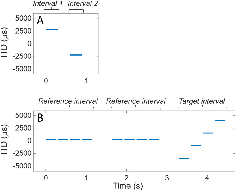
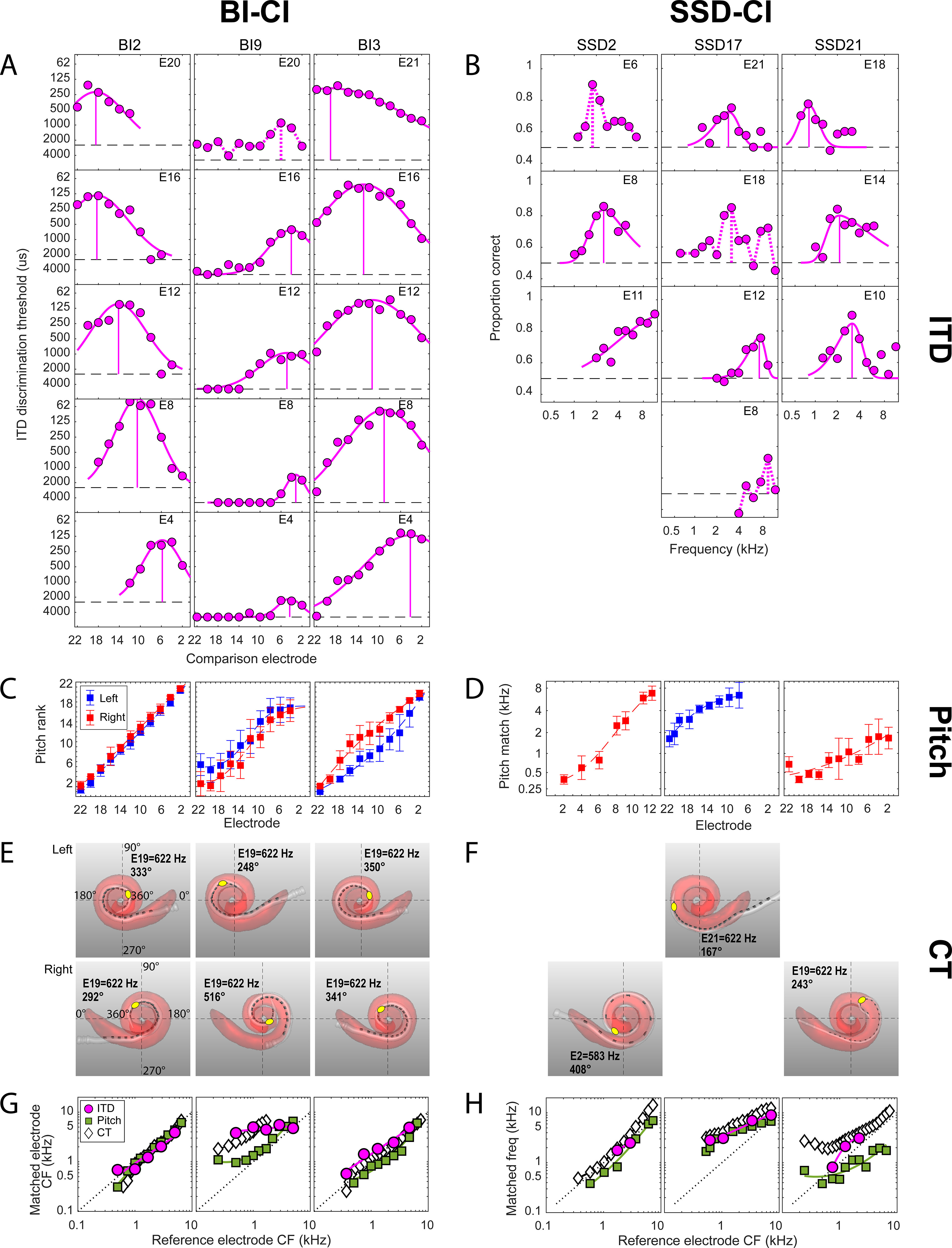
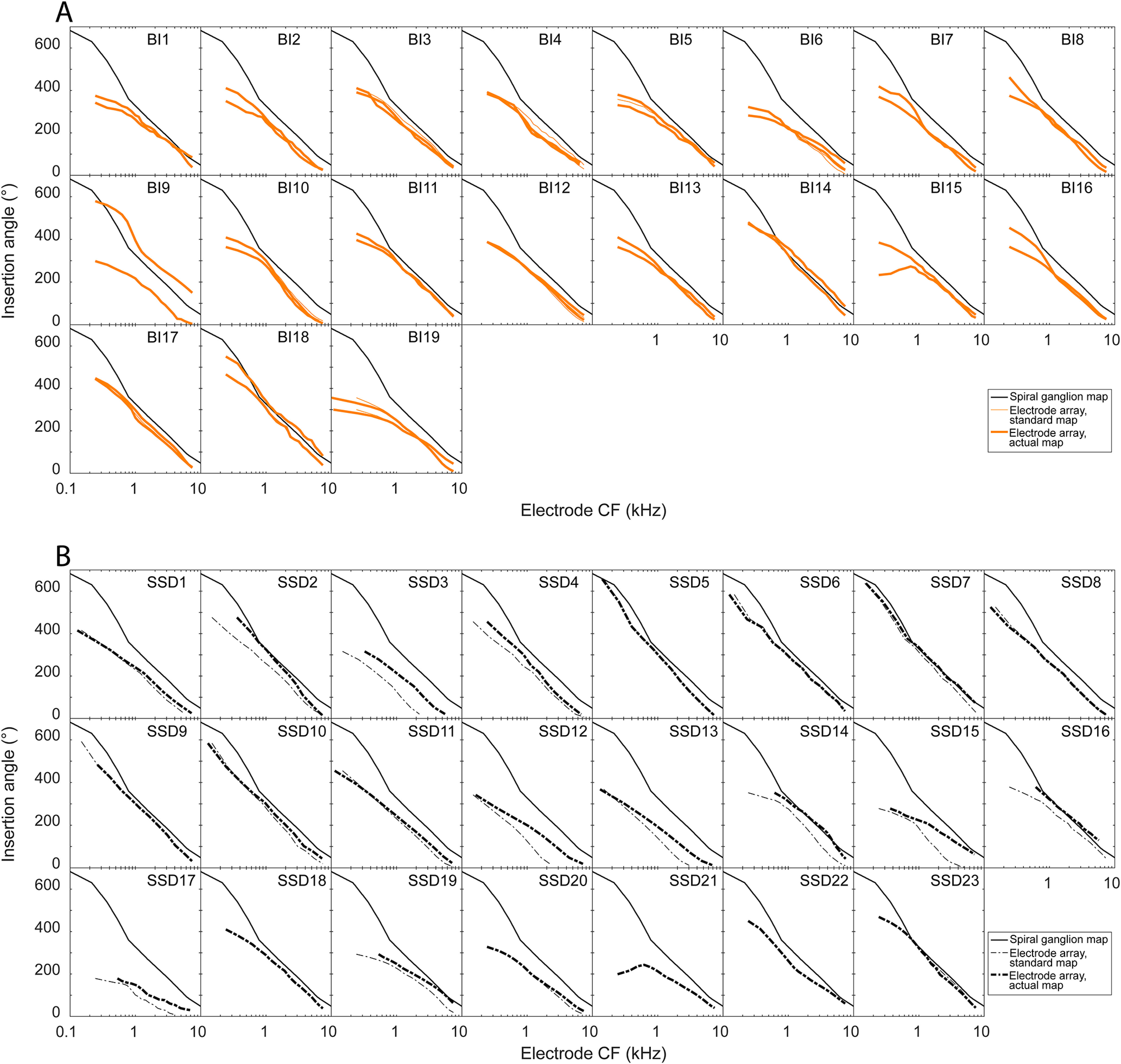
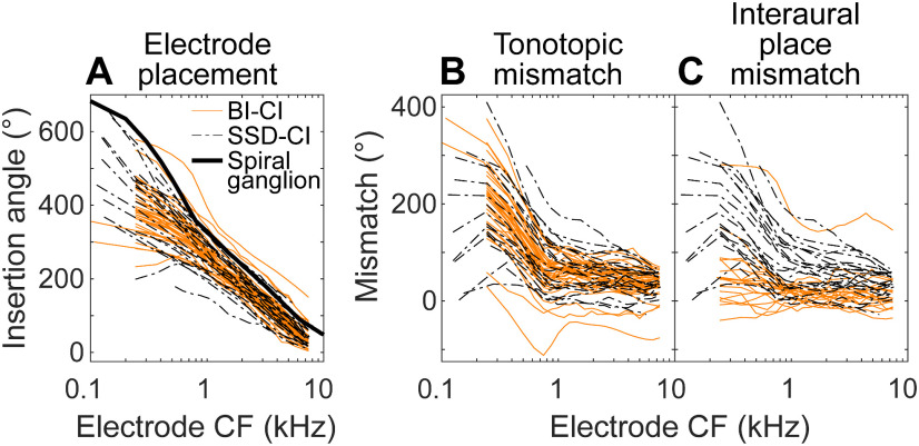
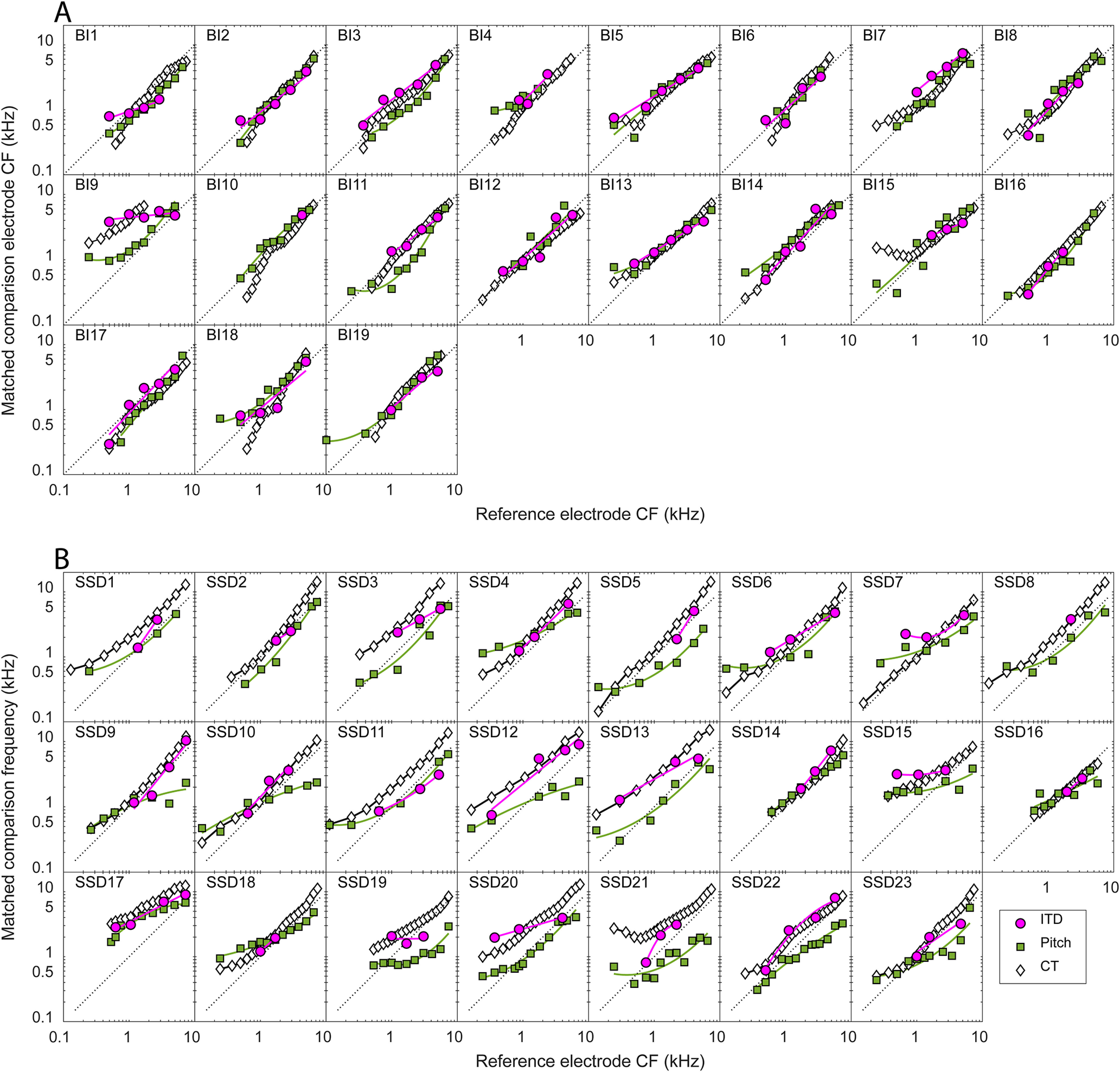
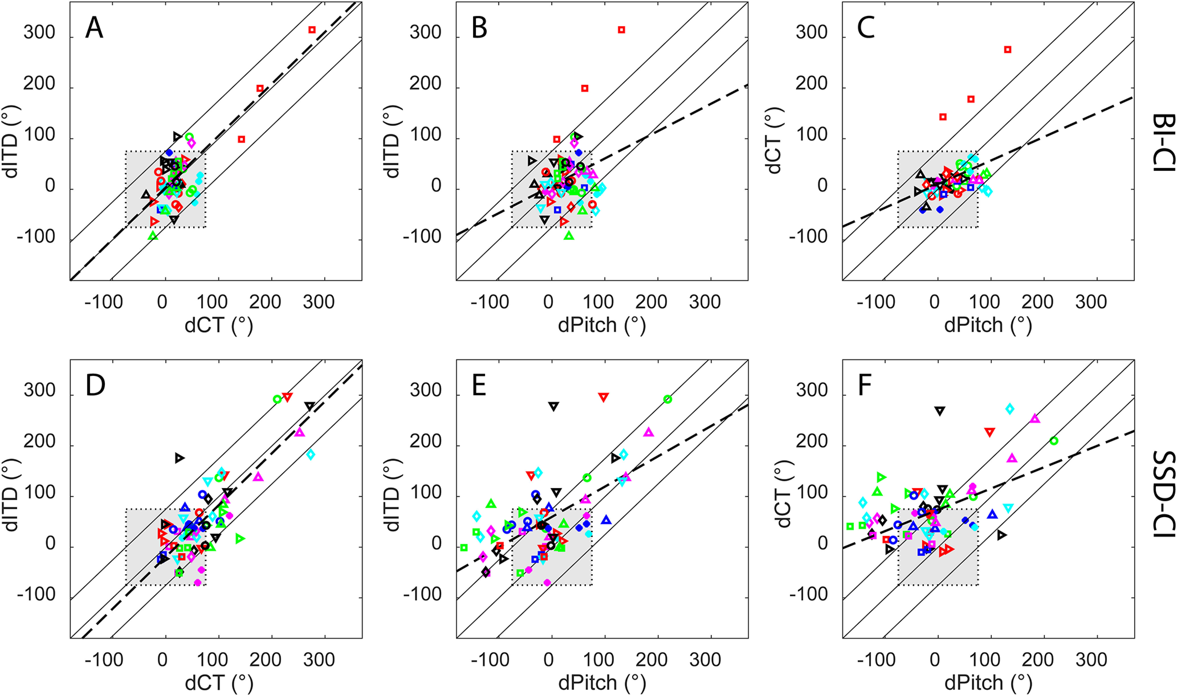
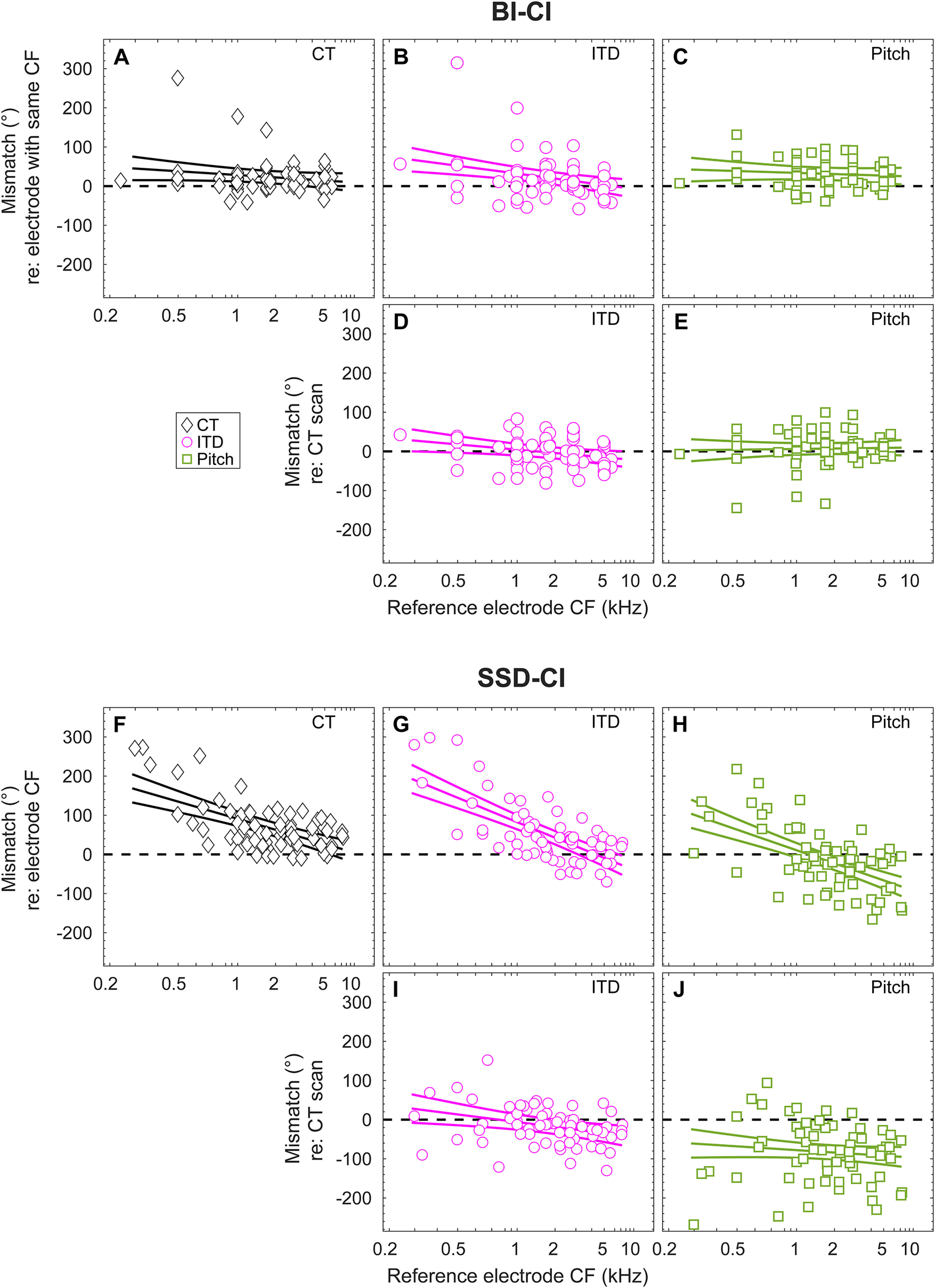
Bilateral cochlear implants (BI-CIs) or a CI for single-sided deafness (SSD-CI; one normally functioning acoustic ear) can partially restore spatial-hearing abilities, including sound localization and speech understanding in noise. For these populations, however, interaural place-of-stimulation mismatch can occur and thus diminish binaural sensitivity that relies on interaurally frequency-matched neurons. This study examined whether plasticity-reorganization of central neural pathways over time-can compensate for peripheral interaural place mismatch. We hypothesized differential plasticity across two systems: none for binaural processing but adaptation for pitch perception toward frequencies delivered by the specific electrodes. Interaural place mismatch was evaluated in 19 BI-CI and 23 SSD-CI human subjects (both sexes) using binaural processing (interaural-time-difference discrimination with simultaneous bilateral stimulation), pitch perception (pitch ranking for single electrodes or acoustic tones with sequential bilateral stimulation), and physical electrode-location estimates from computed-tomography (CT) scans. On average, CT scans revealed relatively little BI-CI interaural place mismatch (26° insertion-angle mismatch) but a relatively large SSD-CI mismatch, particularly at low frequencies (166° for an electrode tuned to 300 Hz, decreasing to 14° at 7000 Hz). For BI-CI subjects, the three metrics were in agreement because there was little mismatch. For SSD-CI subjects, binaural and CT measurements were in agreement, suggesting little binaural-system plasticity induced by mismatch. The pitch measurements disagreed with binaural and CT measurements, suggesting place-pitch plasticity or a procedural bias. These results suggest that reducing interaural place mismatch and potentially improving binaural processing by reprogramming the CI frequency allocation would be better done using CT-scan than pitch information.SIGNIFICANCE STATEMENT Electrode-array placement for cochlear implants (bionic prostheses that partially restore hearing) does not explicitly align neural representations of frequency information. The resulting interaural place-of-stimulation mismatch can diminish spatial-hearing abilities. In this study, adults with two cochlear implants showed reasonable interaural alignment, whereas those with one cochlear implant but normal hearing in the other ear often showed mismatch. In cases of mismatch, binaural sensitivity was best when the same cochlear locations were stimulated in both ears, suggesting that binaural brainstem pathways do not experience plasticity to compensate for mismatch. In contrast, interaurally pitch-matched electrodes deviated from cochlear-location estimates and did not optimize binaural sensitivity. Clinical correction of interaural place mismatch using binaural or computed-tomography (but not pitch) information may improve spatial-hearing benefits.

Keywords: binaural; brainstem; interaural time difference; mismatch; plasticity; superior olivary complex.


Figures

Figure 1.
Figure 1.
Examples of an ITD-discrimination trial. A, For BI-CI subjects, a two-interval, two-alternative forced-choice left/right discrimination task required the subject to determine whether the two stimuli were played left then right or right then left. B, For SSD-CI subjects, the left/right discrimination task proved too difficult given the inherent time delay between the ears. Therefore, a three-interval, two-alternative forced-choice task was used, which required the subject to determine whether the second or third interval contained a moving set of stimuli.
Figure 2.
Figure 2.
Examples of the perceptual and objective data collected for the three interaural place-mismatch estimation methods for six example subjects (3 BI-CI and 3 SSD-CI). For each subject, the reference ear is defined as the functionally poorer ear as determined by speech understanding scores. A, B, ITD discrimination data. Discrimination threshold (BI-CI subjects) or proportion correct (SSD-CI subjects) as a function of the comparison electrode or frequency for a given reference electrode. The dashed horizontal line shows where threshold could not be measured (A) or chance performance (B). The vertical line shows the estimated interaural place match. C, D, Pitch ranking data; rank (BI-CI subjects) or pitch match (SSD-CI subjects) as a function of electrode number. Error bars indicate the standard deviation of the rank or match. E, F, Computational model output of the CT scan analysis, showing the estimated cochlear morphology (red), the position of the electrode array (white), and the individual electrode contacts (dark gray). G, H, Summary of the three estimates of interaural place match as a function of reference electrode number (BI-CI subjects) or reference electrode CF (SSD-CI subjects) derived from the measurements in A–F. The diagonal dotted lines in G and H represent perfect interaural alignment; vertical displacement from this line represents mismatch.
Figure 3.
Figure 3.
CT scan data for each individual subject in the study showing the absolute insertion angles of each electrode in the array for the BI-CI subjects (A) and the SSD-CI subjects (B). The thin lines plot the insertion depth as a function of the specific default frequency allocation for each electrode from the manufacturer. The thick lines plot the insertion angle as a function of the subject's actual frequency allocation, which differs in some cases from the default of the manufacturer. The solid black line is an absolute reference line depicting the relationship between the electrode insertion angle and the spiral ganglion characteristic frequency at that location (Stakhovskaya et al., 2007).
Figure 4.
Figure 4.
A, Comparison of the absolute electrode positions for the BI-CI (orange) and SSD-CI subjects (black) as a function of the actual frequency allocation of the electrode. B, The tonotopic mismatch between the insertion angle and the electrode frequency allocation was similar for the two groups. C, The interaural place mismatch, which reflects the relationship between the two electrode insertions for the BI-CI subjects but reflects the tonotopic place mismatch for the SSD-CI subjects, was smaller for the BI-CI subjects.
Figure 5.
Figure 5.
Summary of the three estimates of relative interaural place for each individual subject in the study. A, For the BI-CI subjects, the matched electrode in the comparison ear is plotted as a function of the reference electrode. B, For the SSD-CI subjects, the matched acoustic frequency in the comparison ear is plotted as a function of the reference electrode CF. The diagonal dotted lines represent perfect interaural alignment; vertical displacement from this line represents mismatch.
Figure 6.
Figure 6.
Pairwise comparisons of the three estimates of the magnitude of interaural place mismatch for individual electrodes for A–C, the BI-CI subjects, and D–F, the SSD-CI subjects, with each color/symbol combination representing a different subject. The three diagonal lines indicate 1:1 correspondence (±75°) where two mismatch estimates are not different. The gray shaded box indicates the ±75° mismatch range (equivalent to 3 mm on the Greenwood scale) over which binaural sensitivity is estimated to be tolerant to mismatch (Kan et al., 2013). The dashed lines represent linear fits from a regression analysis.
Figure 7.
Figure 7.
Tonotopic dependence of interaural place mismatch for A–E, BI-CI subjects, and F–J, SSD-CI subjects. The first and third rows reference the mismatch to the clinical map, based on A–C, the insertion angle associated with the number-matched electrode for BI-CI subjects, or F–H, the insertion angle associated with the reference electrode CF for SSD-CI subjects. The second and fourth rows (D, E and I, J) reference the mismatch to the CT estimate of electrode position. Points represent mismatch measurements for individual electrodes; fitted curves represent estimates of the group average and 95% confidence interval. Horizontal dashed lines indicate zero mismatch.

References

    1. Adel Y, Nagel S, Weissgerber T, Baumann U, Macherey O (2019) Pitch matching in cochlear implant users with single-sided deafness: effects of electrode position and acoustic stimulus type. Front Neurosci 13:1119. - PMC - PubMed
    1. Aronoff JM, Padilla M, Stelmach J, Landsberger DM (2016) Clinically paired electrodes are often not perceived as pitch matched. Trends Hear 20:233121651666830. 10.1177/2331216516668302 - DOI - PMC - PubMed
    1. Aronoff JM, Staisloff HE, Kirchner A, Lee DH, Stelmach J (2019) Pitch matching adapts even for bilateral cochlear implant users with relatively small initial pitch differences across the ears. J Assoc Res Otolaryngol 20:595–603. 10.1007/s10162-019-00733-3 - DOI - PMC - PubMed
    1. Bernstein JGW, Oxenham AJ (2003) Pitch discrimination of diotic and dichotic tone complexes: harmonic resolvability or harmonic number? J Acoust Soc Am 113:3323–3334. 10.1121/1.1572146 - DOI - PubMed
    1. Bernstein JGW, Goupell MJ, Schuchman GI, Rivera AL, Brungart DS (2016) Having two ears facilitates the perceptual separation of concurrent talkers for bilateral and single-sided deaf cochlear implantees. Ear Hear 37:289–302. 10.1097/AUD.0000000000000284 - DOI - PMC - PubMed

Publication types