

A comparative analysis of cell surface targeting aptamers
- PMID: 34725326
- PMCID: PMC8560833
- DOI: 10.1038/s41467-021-26463-w
A comparative analysis of cell surface targeting aptamers
Abstract
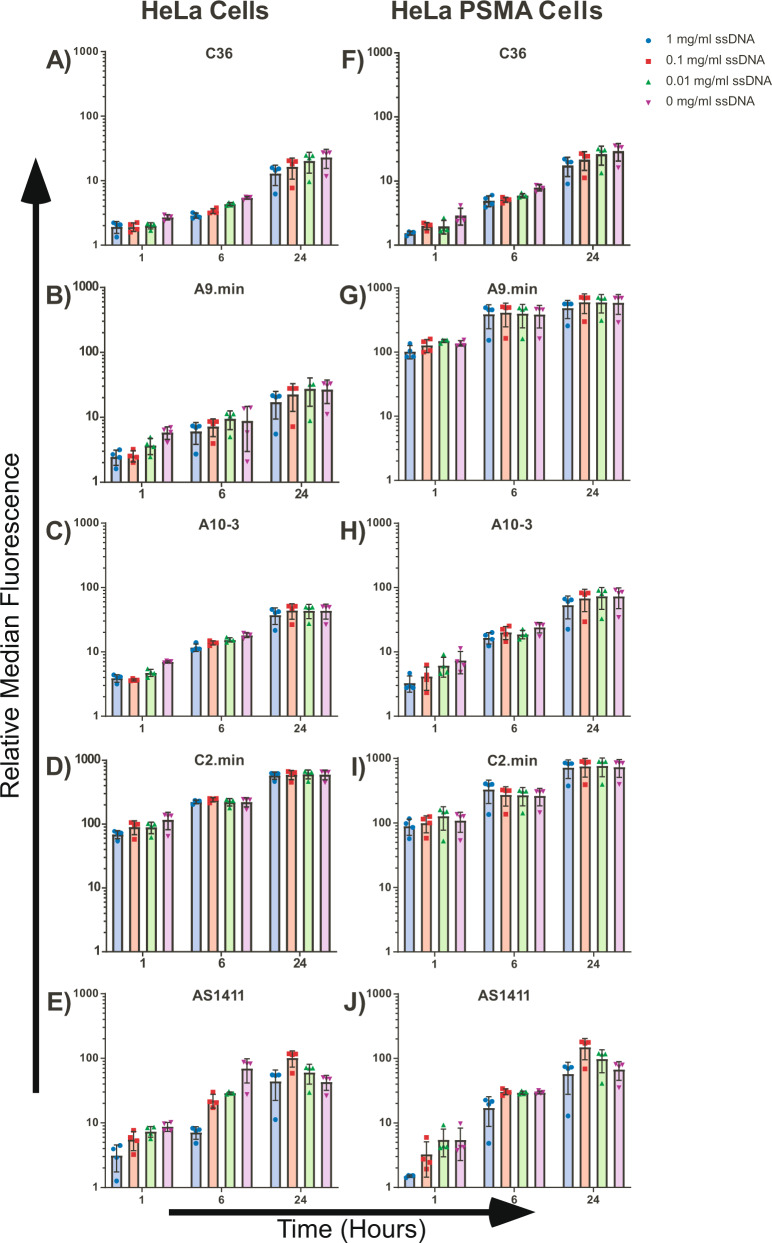
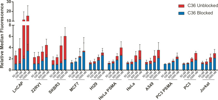
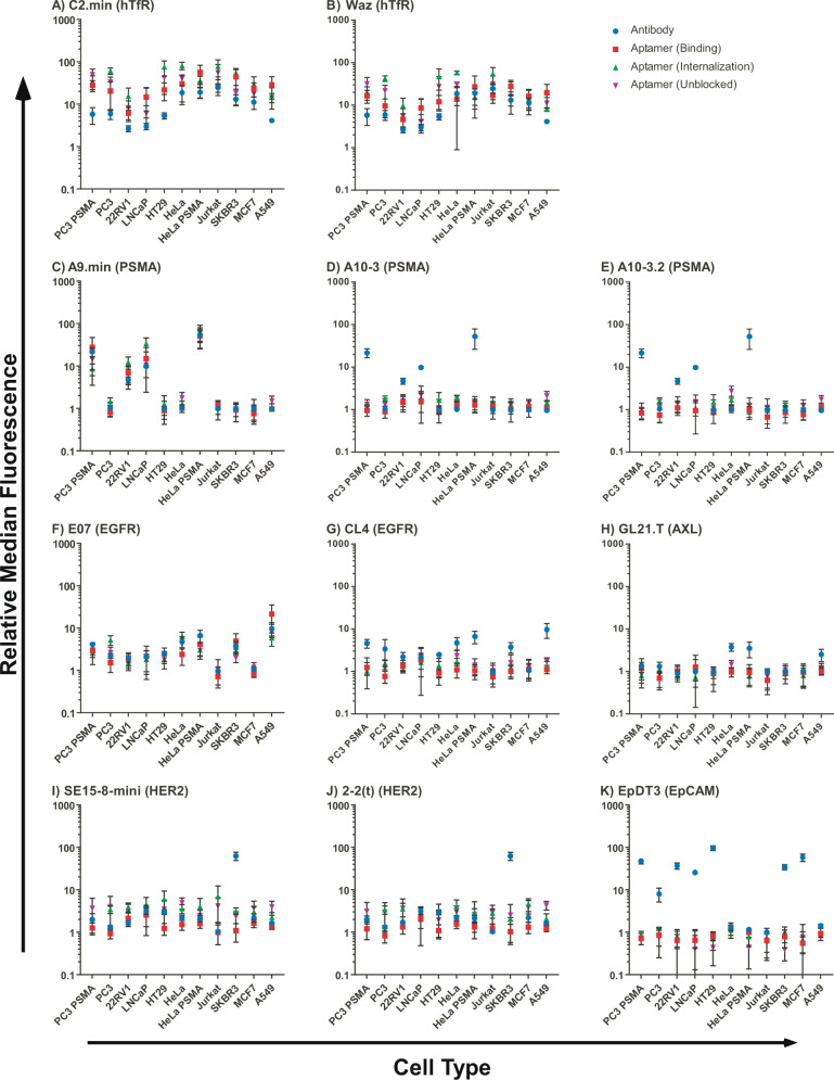
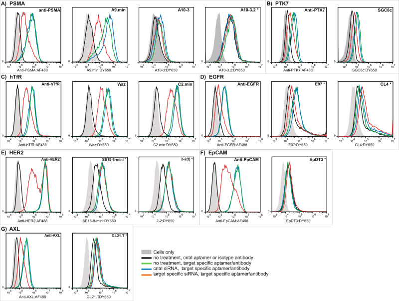
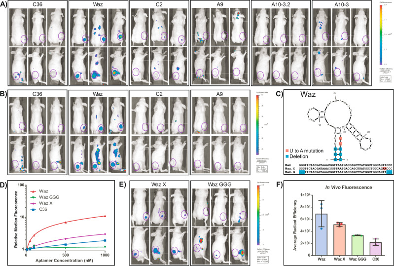
Aptamers represent a potentially important class of ligands for the development of diagnostics and therapeutics. However, it is often difficult to compare the function and specificity of many of these molecules as assay formats and conditions vary greatly. Here, with an interest in developing aptamer targeted therapeutics that could effectively deliver cargoes to cells, we chemically synthesize 15 aptamers that have been reported to target cell surface receptors or cells. Using standardized assay conditions, we assess each aptamer's binding properties on a panel of 11 different cancer cell lines, correlate aptamer binding to antibody controls and use siRNA transfection to validate each aptamer's binding to reported target receptors. Using a subset of these molecules known to be expressed on prostate cancers, we use near-infrared in vivo imaging to assess the tumor localization following intravenous injection. Our data demonstrate some surprising differences in the reported specificity and function for many of these molecules and raise concerns regarding their cell targeting capabilities. They also identify an anti-human transferrin aptamer, Waz, as a robust candidate for targeting prostate cancers and for future development of aptamer-based therapeutics.
© 2021. The Author(s).
Conflict of interest statement
The authors declare no competing interests.
Figures
Similar articles
-
Aptamer-Mediated Delivery and Cell-Targeting Aptamers: Room for Improvement.Nucleic Acid Ther. 2018 Jun;28(3):194-199. doi: 10.1089/nat.2018.0732. Nucleic Acid Ther. 2018. PMID: 29883295 Free PMC article. Review.
-
Development of cell-type specific anti-HIV gp120 aptamers for siRNA delivery.J Vis Exp. 2011 Jun 23;(52):2954. doi: 10.3791/2954. J Vis Exp. 2011. PMID: 21730942 Free PMC article.
-
G-Quadruplex-Proximized Aptamers (G4PA) Efficiently Targeting Cell-Surface Transferrin Receptors for Targeted Cargo Delivery.Nano Lett. 2022 Aug 10;22(15):6328-6333. doi: 10.1021/acs.nanolett.2c02064. Epub 2022 Jul 28. Nano Lett. 2022. PMID: 35900277
-
From ugly duckling to swan: unexpected identification from cell-SELEX of an anti-Annexin A2 aptamer targeting tumors.PLoS One. 2014 Jan 29;9(1):e87002. doi: 10.1371/journal.pone.0087002. eCollection 2014. PLoS One. 2014. PMID: 24489826 Free PMC article.
-
Chimeric aptamers in cancer cell-targeted drug delivery.Crit Rev Biochem Mol Biol. 2011 Dec;46(6):459-77. doi: 10.3109/10409238.2011.614592. Epub 2011 Sep 28. Crit Rev Biochem Mol Biol. 2011. PMID: 21955150 Free PMC article. Review.
Cited by
-
Targeting triple-negative breast cancer cells with a β1-integrin binding aptamer.Mol Ther Nucleic Acids. 2023 Aug 16;33:871-884. doi: 10.1016/j.omtn.2023.08.015. eCollection 2023 Sep 12. Mol Ther Nucleic Acids. 2023. PMID: 37680989 Free PMC article.
-
Anti-EGFR aptamer exhibits direct anti-cancer effects in NSCLC cells harboring EGFR L858R mutations.NPJ Precis Oncol. 2024 Nov 21;8(1):271. doi: 10.1038/s41698-024-00758-9. NPJ Precis Oncol. 2024. PMID: 39572699 Free PMC article.
-
Anti-IL-4Rα aptamer reduces IL-4 signalling and formation of nasal polyps.ERJ Open Res. 2025 Jun 16;11(3):00700-2024. doi: 10.1183/23120541.00700-2024. eCollection 2025 May. ERJ Open Res. 2025. PMID: 40524931 Free PMC article.
-
Aptamer-Targeted Dendrimersomes Assembled from Azido-Modified Janus Dendrimers "Clicked" to DNA.Biomacromolecules. 2024 Mar 11;25(3):1541-1549. doi: 10.1021/acs.biomac.3c01108. Epub 2024 Feb 23. Biomacromolecules. 2024. PMID: 38394608 Free PMC article.
-
Aptamers: Design, Theory, and Applications to Diagnosis and Therapy for Diseases.MedComm (2020). 2025 May 19;6(5):e70180. doi: 10.1002/mco2.70180. eCollection 2025 May. MedComm (2020). 2025. PMID: 40391083 Free PMC article. Review.
References
-
- Bradbury A, Pluckthun A. Standardize antibodies used in research: to save millions of dollars and dramatically improve reproducibility, protein-binding reagents must be defined by their sequences and produced as recombinant proteins, say Andrew Bradbury, Andreas Pluckthun and 110 co-signatories. Nature. 2015;518:27–30. doi: 10.1038/518027a. - DOI - PubMed
Publication types
MeSH terms
Substances
Grants and funding
LinkOut - more resources
Full Text Sources
Other Literature Sources
Medical
