

A novel method quantifying caries following orthodontic treatment
- PMID: 34725354
- PMCID: PMC8560919
- DOI: 10.1038/s41598-021-00561-7
A novel method quantifying caries following orthodontic treatment
Abstract
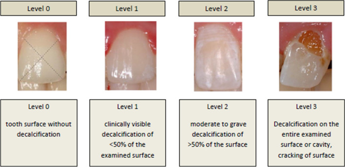
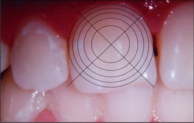
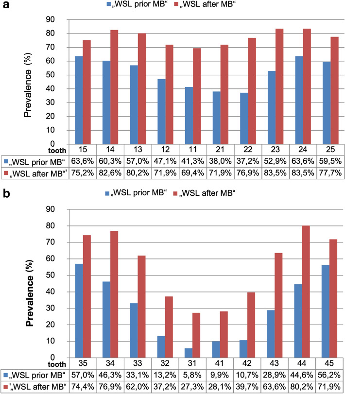
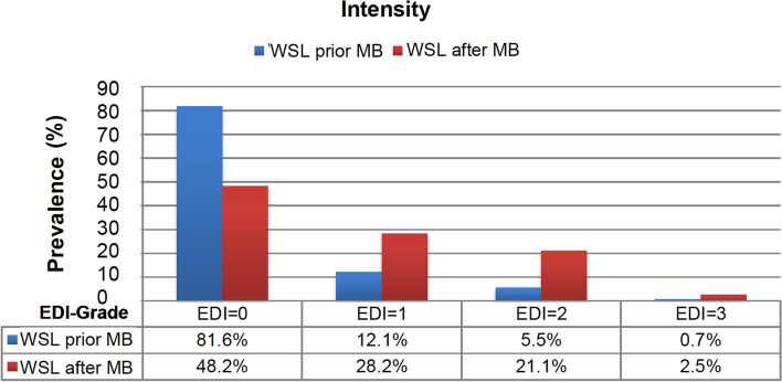
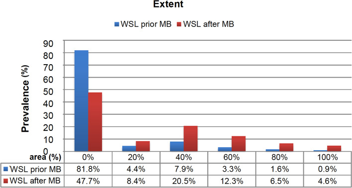
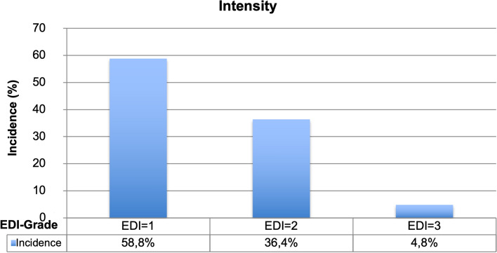
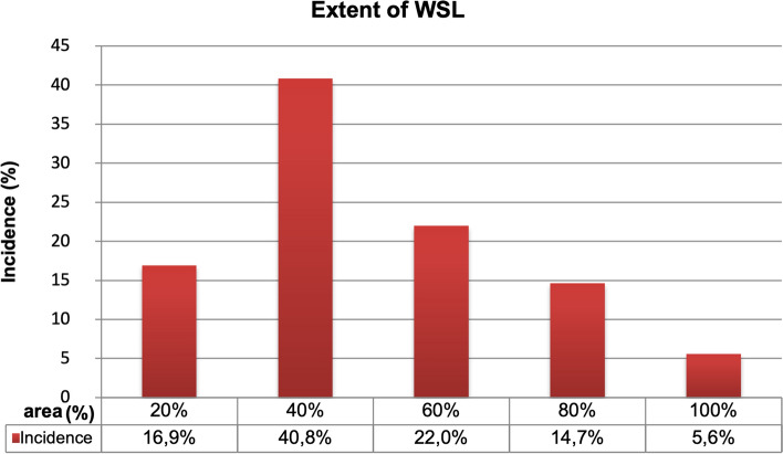
This retrospective pilot study used a newly developed evaluation tool to assess the prevalence and incidence of White Spot Lesions (WSL) before and after multibracket appliance (MB) therapy. Digital photographs of 121 adolescent patients (63 ♂, 58 ♀) with metal brackets were analyzed retrospectively before and after MB therapy. The labial surfaces of anterior teeth, canine teeth, and premolars in the upper (UJ) and lower jaws (LJ) were evaluated using the Enamel Decalcification Index (EDI) by Banks and Richmond (Eur J Orthod, 16(1):19-25, 1994, levels 0-3) and a specially developed digitally scaled graticule with concentric circles to quantify the extent of WSL (in %). The statistical data analysis was based on crosstabulations and logistic regression. Before MB, 69.4% of the patients presented at least one WSL and 97.5% after, an increase of 28.1%. Before MB, 18.4% of the tooth surfaces (TS) showed an EDI level of 1-3. After MB, 51.8% of the TS featured WSL. 18.2% of the TS showed a WSL to the extent of ≥ 20-100% before and 52.3% after MB. The incidence in the UJ (71-79%) as well as the LJ (64-76%) was highest for the first and second premolars and lowest for LJ incisors (22-35%). The probability for developing a new distal WSL is higher than developing gingival, mesial or occlusal WSL. Labial MB therapy drastically increases the risk of developing WSL. We verified a concise quantification of the extent of labial WSL with the evaluation index.
© 2021. The Author(s).
Conflict of interest statement
The authors declare no competing interests.
Figures
References
-
- Micheelis W, Schiffner U. Vierte Deutsche Mundgesundheitsstudie (DMS IV) Ärzte Verlag; 2006.
-
- Ogaard B. Cariologic aspects of orthodontic treatment. Nor. Tannlaegeforen Tid. 1989;99(20):802–805. - PubMed
Publication types
MeSH terms
LinkOut - more resources
Full Text Sources
Medical
Miscellaneous
