. 2021 Nov 1;4(1):1237.
doi: 10.1038/s42003-021-02744-4.

EGFR Regulates the Hippo pathway by promoting the tyrosine phosphorylation of MOB1

Affiliations

EGFR Regulates the Hippo pathway by promoting the tyrosine phosphorylation of MOB1

Toshinori Ando et al. Commun Biol. .

Abstract

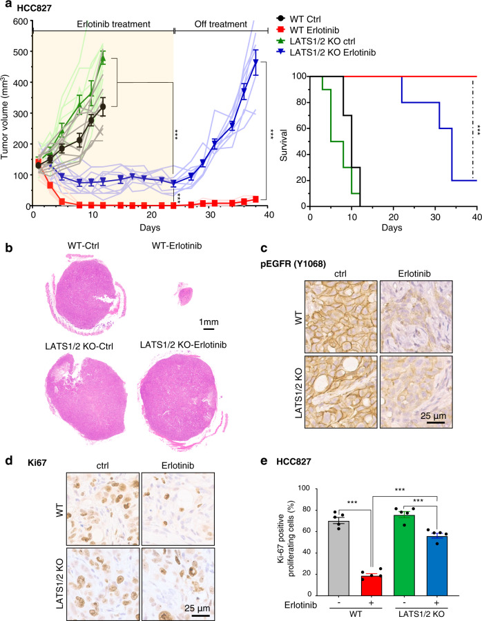
The Hippo pathway is frequently dysregulated in cancer, leading to the unrestrained activity of its downstream targets, YAP/TAZ, and aberrant tumor growth. However, the precise mechanisms leading to YAP/TAZ activation in most cancers is still poorly understood. Analysis of large tissue collections revealed YAP activation in most head and neck squamous cell carcinoma (HNSCC), but only 29.8% of HNSCC cases present genetic alterations in the FAT1 tumor suppressor gene that may underlie persistent YAP signaling. EGFR is overexpressed in HNSCC and many other cancers, but whether EGFR controls YAP activation is still poorly understood. Here, we discover that EGFR activates YAP/TAZ in HNSCC cells, but independently of its typical signaling targets, including PI3K. Mechanistically, we find that EGFR promotes the phosphorylation of MOB1, a core Hippo pathway component, and the inactivation of LATS1/2 independently of MST1/2. Transcriptomic analysis reveals that erlotinib, a clinical EGFR inhibitor, inactivates YAP/TAZ. Remarkably, loss of LATS1/2, resulting in aberrant YAP/TAZ activity, confers erlotinib resistance on HNSCC and lung cancer cells. Our findings suggest that EGFR-YAP/TAZ signaling plays a growth-promoting role in cancers harboring EGFR alterations, and that inhibition of YAP/TAZ in combination with EGFR might be beneficial to prevent treatment resistance and cancer recurrence.


Conflict of interest statement

The authors declare the following competing interests: J.S.G. is a member of the advisory board of Oncoceutics, Domain Therapeutics, and Vividion. The remaining authors declare no competing interests.

Figures

Fig. 1
Fig. 1. EGFR activates YAP/TAZ in HNSCC, independently of PI3K.
a Immunoblot of pEGFR (Y1068), EGFR, pYAP (S127), YAP, pERK1/2 (T202/Y204), ERK1/2, pAKT (S473), AKT, pS6 (S235/236), S6, CTGF, CYR61, β-actin in CAL33, CAL27, and WSU-HN6. Right panel showing the status of FAT1 gene alterations. b Relative mRNA levels of CTGF, CYR61, and AMOTL2 in CAL33, CAL27, and WSU-HN6 cells. c GSEA analysis of RNA-seq data in CCLE using the C6 oncogenic gene sets, spiked with several previously published YAP-regulated gene sets. NES, normalized enrichment score; NOM, nominal; FDR, false discovery rate. d Immunoblot of pEGFR (Y1068), EGFR, pYAP (S127), YAP, TAZ, pERK1/2 (T202/Y204), ERK1/2, pAKT (S473), AKT, pS6 (S235/236), S6, CTGF, CYR61, β-actin in CAL27 cells. Cells were serum starved for 16 h, and treated with EGF (20 ng/ml) for the indicated time. e Relative mRNA levels of CTGF, CYR61, and AMOTL2 in CAL27 cells. f Immunoblot of pEGFR (Y1068), EGFR, pAKT (S473), AKT, pYAP (S127), YAP, TAZ, CTGF, CYR61, β-actin in CAL27 cells stably overexpressing empty vector or PIK3CA H1047R. Cells were serum starved for 16 h, and treated with EGF (20 ng/ml) for 1 hr. g Immunoblot of pEGFR (Y1068), EGFR, pAKT (S473), AKT, pYAP (S127), YAP, TAZ, CTGF, CYR61, β-actin in CAL27 cells. Cells were serum starved for 16 h, and pretreated with BYL719 (1 μM) for 1 h and followed by EGF treatment (20 ng/ml) for 1 h. ANOVA with Tukey–Kramer post hoc test was used. Mean ± SEM (b, e); ***P  < 0.001; **P < 0.01; *P  < 0.05. *versus CAL33 (b) and versus EGF 0 h (e).
Fig. 2
Fig. 2. EGFR under-phosphorylates YAP/TAZ, induces nuclear translocation of YAP/TAZ and their interaction with TEADs, promoting CTGF/CYR61/AMOTL2 expression.
a Immunoblot of EGFR, pEGFR (Y1068), pYAP (S127), YAP, TAZ, pERK1/2 (T202/Y204), ERK1/2, pAKT (S473), AKT, pS6 (S235/236), S6, CTGF, CYR61, β-actin in vector- or EGFR-overexpressing HEK293A cells. Cells were serum starved for 16 h, and treated with EGF (20 ng/ml) for the indicated time. b Relative mRNA levels of CTGF, CYR61, and AMOTL2. c Immunoblot of pEGFR (Y1068), EGFR, pYAP (S127), YAP, TAZ, CTGF, CYR61, β-actin in EGFR-overexpressing HEK293A cells. Cells were transfected with siRNA control and against YAP/TAZ for 24 h, serum starved for 16 h, and treated with EGF (20 ng/ml) for 1 h. d Relative mRNA levels of CTGF, CYR61, and AMOTL2. e Co-immunoprecipitation of YAP and TEAD1. Lysates were immunoprecipitated with control IgG or an antibody against YAP. Immunoblot of TEAD1, YAP, pYAP (S127), pEGFR (Y1068), EGFR, β-actin in EGFR-overexpressing HEK293A cells. Cells were serum stared for 16 h, and treated with EGF (20 ng/ml) for 1 h. f YAP/TAZ localization analyzed by immunofluorescence staining. Cells were serum starved for 16 h, and treated with EGF (20 ng/ml) for 1 h. Scale bars indicate 5 μm. ANOVA with Tukey–Kramer post hoc test was used. Mean ± SEM (b, d); ***P <0.001; **P <0.01; *P <0.05. *versus EGF 0 h (b).
Fig. 3
Fig. 3. EGFR stimulation leads to MOB1 phosphorylation and LATS1/2 inactivation, independently of MST1/2.
a Immunoprecipitation of LATS1. Lysates were immunoprecipitated with control IgG or an antibody against LATS1. Immunoblot of pLATS1 (T1079), LATS1, pEGFR (Y1068), EGFR, β-actin in EGFR-overexpressing HEK293A cells. Cells were serum stared for 16 h, and treated with EGF (20 ng/ml) for 1 h. b In vitro kinase assay of LATS1 against YAP. Lysates were immunoprecipitated with control IgG or an antibody against LATS1, then applied for in vitro kinase reaction with GST-YAP protein. Cells were serum starved for 16 h, and treated with EGF (20 ng/ml) or FBS (10%) as positive control for 1 h. Immunoblot of pYAP (S127), GST, LATS1, pEGFR (Y1068), EGFR, β-actin. Arrow indicates non-specific band. c Immunoblot of LATS1, LATS2, pEGFR (Y1068), EGFR, pYAP (S127), YAP, TAZ, β-actin in WT, or LATS1/2 KO HEK293A cells. WT or LATS1/2 KO HEK293A cells were transfected with EGFR plasmid and incubated for 24 h, serum starved for 16 h, and treated with EGF (20 ng/ml) for 1 h. d Immunoprecipitation of Myc-MST1, FLAG-SAV1, FLAG-LATS1, HA-MOB1, and Myc-YAP. Lysates were immunoprecipitated with control IgG or antibodies against each Tag. Immunoblot of pY, Tag, pEGFR (Y1068), EGFR, β-actin. EGFR-overexpressing HEK293A cells were transfected with the Hippo-components and YAP plasmid, incubated for 24 h, serum starved for 16 h, and treated with EGF (20 ng/ml) for 1 h. e In vitro kinase assay of EGFR and GST-MOB1. In vitro kinase reaction was performed with recombinant EGFR, GST-MOB1 protein, and ATP. Immunoblot for pY and GST. f Co-immunoprecipitation of HA-MOB1 and LATS1. Lysates were immunoprecipitated with control IgG or an antibody against HA-tag. Immunoblot of LATS1, HA, pEGFR (Y1068), EGFR, β-actin. EGFR-overexpressing HEK293A cells were transfected with HA-MOB1 plasmid and incubated for 24 h, serum starved for 16 h, and treated with EGF (20 ng/ml) for 1 h. g Immunoblot of Myc-tag, pMST1/2 (T183/T180), pMOB1 (T35), MOB1, pEGFR (Y1068), EGFR, β-actin. EGFR-overexpressing HEK293A cells were transfected with vector or Myc-MST1 plasmid and incubated for 24 h, serum starved for 16 h, and treated with EGF (20 ng/ml) for 1 h. Asterisks indicate non-specific bands.
Fig. 4
Fig. 4. EGFR activation leads to MOB1 phosphorylation at Y95, 114, 117.
a Immunoprecipitation of HA-MOB1 WT and 8YF. Lysates were immunoprecipitated with an antibody against HA-tag. Immunoblot of pY, HA, pEGFR (Y1068), EGFR, β-actin. EGFR-overexpressing HEK293A cells were transfected with HA-MOB1 WT or 8YF plasmid and incubated for 24 h, serum starved for 16 h, and treated with EGF (20 ng/ml) for 1 h. b Schematic of “Add-back approach”. All 8 tyrosines of MOB1 WT were mutated into phenylalanine (8YF), then each site was mutated back to tyrosine (7YF + Y). c Immunoprecipitation of HA-MOB1 WT, 8YF and 7YF + Y26, Y72, Y93, Y95, Y114, Y117, Y159, Y163. Lysates were immunoprecipitated with an antibody against HA-tag. Immunoblot of pY, HA, pEGFR (Y1068), EGFR, β-actin. EGFR-overexpressing HEK293A cells were transfected with HA-MOB1 WT, 8YF, and 7YF + Y mutant plasmid and incubated for 24 h, serum starved for 16 h, and treated with EGF (20 ng/ml) for 1 h. d The conserved amino acid sequences at Y95, Y114, Y117 of MOB1A in various species. e Immunoprecipitation of HA-MOB1 WT and 3YF. Lysates were immunoprecipitated with an antibody against HA-tag. Immunoblot of pY, HA, pEGFR (Y1068), EGFR, β-actin. EGFR-overexpressing HEK293A cells were transfected with HA-MOB1 WT or 3YF plasmid and incubated for 24 h, serum starved for 16 h, and treated with EGF (20 ng/ml) for 1 h. f Relative mRNA expression of CTGF. ANOVA with Tukey–Kramer post hoc test was used. Mean ± SEM (f); ***P <0.001; **P <0.01.
Fig. 5
Fig. 5. Erlotinib increases pYAP and suppresses YAP/TAZ-regulated genes.
a Immunoblot of pEGFR (Y1068), EGFR, pYAP (S127), YAP, β-actin in WSU-HN6 and HCC827 cells. Cells were treated with erlotinib at the indicated concentrations for 2 h. b Relative mRNA levels of CTGF, CYR61, and AMOTL2 in WSU-HN6 and HCC827 cells. Cells were treated with erlotinib (1 μM) for 2 h. c The top 15 enriched oncogenic signatures gene sets from RNA-seq data analysis of HCC827 cells. YAP-regulated signatures gene sets are highlighted in red. Cells were treated with vehicle or erlotinib (1 μM) for 24 h. The original name of signature gene sets are listed in supplementary Fig. S5a. d Heat map showing Z-score normalized mRNA expression of representative YAP/TAZ-regulated genes highlighted in orange and yellow. The genes highlighted in green and blue are consistent with the ones previously reported as up- or downregulated by erlotinib treatment. e Enrichment plots of YAP conserved signatures. f Immunoblot of YAP, TAZ, β-actin in HN6 and HCC827 cells. Cells were transfected with siRNA for control and YAP/TAZ, and incubated for 48 h. g Cell viability. ANOVA with Tukey–Kramer post hoc test and Student’s t-test were used. Mean ± SEM (b, g); ***P <0.001; **P <0.01.
Fig. 6
Fig. 6. Loss of LATS1/2 confers resistance to erlotinib treatment in cancer cells with EGFR alterations.
a Immunoblot of LATS1, LATS2, pYAP (S127), YAP, TAZ, β-actin in WT or LATS1/2 KO HN6 and HCC827 cells. b Relative mRNA levels of CTGF, CYR61, and AMOTL2. Cells were treated with erlotinib (1 μM) for 2 h. c Cell viability. Cells were treated with erlotinib for 3 days. d Schematic of EGFR-mediated YAP/TAZ activation. When EGFR is inactivated, the hydrophobic site of LATS1/2 is phosphorylated and LATS1/2 are active, leading to YAP/TAZ phosphorylation and cytoplasmic retention or degradation. Upon EGF stimulation or EGFR activation by gene amplification, overexpression or mutations, MOB1 is tyrosine phosphorylated and LATS1/2 are dephosphorylated and inactive, resulting in YAP/TAZ nuclear translocation and expression of growth promoting genes regulated by YAP/TAZ. ANOVA with Tukey–Kramer post hoc test were used. Mean ± SEM (b); ***P <0.001; **P <0.01. *versus WT erlotinib non-treated.
Fig. 7
Fig. 7. Loss of LATS1/2 confers resistance to erlotinib treatment in cancer cells with EGFR alterations in vivo.
a (left) Individual and average (bold line) growth curves for HCC827 control and LATS1/2 KO cells transplanted into Female NOD-scid IL2Rgammanull mice and treated with erlotinib for 24 days. (n = 10 per group). Tumor re-growth was monitored after erlotinib treatment was discontinued. (right) Kaplan–Meier curve showing survival of mice from (a). The death of animals occurred either naturally, when tumor growth compromised animal welfare, or when tumor volume reached >200% of initial size at day 1 of treatment. (n = 10 mice per group; Log-Rank/Mantel–Cox test.). b Representative histological sections from each treatment group. Scale bar represents 1 mm. c, d Representative immunohistochemical analysis of pEGFR and Ki67 in the short-term treatment groups (every day for 5 days). e Quantification of (d) showing percentage of cells staining positive for Ki67 (n = 5 mice per group). Mean±SEM (a, e); ***P < 0.001.

References

    1. Yu FX, Zhao B, Guan KL. Hippo pathway in organ size control, tissue homeostasis, and cancer. Cell. 2015;163:811–828. doi: 10.1016/j.cell.2015.10.044. - DOI - PMC - PubMed
    1. Zhao B, Li L, Lei Q, Guan KL. The Hippo-YAP pathway in organ size control and tumorigenesis: an updated version. Genes Dev. 2010;24:862–874. doi: 10.1101/gad.1909210. - DOI - PMC - PubMed
    1. Zhao B, Li L, Tumaneng K, Wang CY, Guan KL. A coordinated phosphorylation by Lats and CK1 regulates YAP stability through SCF(beta-TRCP) Genes Dev. 2010;24:72–85. doi: 10.1101/gad.1843810. - DOI - PMC - PubMed
    1. Moroishi T, Hansen CG, Guan KL. The emerging roles of YAP and TAZ in cancer. Nat. Rev. Cancer. 2015;15:73–79. doi: 10.1038/nrc3876. - DOI - PMC - PubMed
    1. Plouffe SW, Hong AW, Guan KL. Disease implications of the Hippo/YAP pathway. Trends Mol. Med. 2015;21:212–222. doi: 10.1016/j.molmed.2015.01.003. - DOI - PMC - PubMed

Publication types