. 2021 Nov;5(11):1261-1273.
doi: 10.1038/s41551-021-00800-2. Epub 2021 Nov 1.

Rejuvenation of tumour-specific T cells through bispecific antibodies targeting PD-L1 on dendritic cells

Affiliations

Rejuvenation of tumour-specific T cells through bispecific antibodies targeting PD-L1 on dendritic cells

Longchao Liu et al. Nat Biomed Eng. 2021 Nov.

Abstract

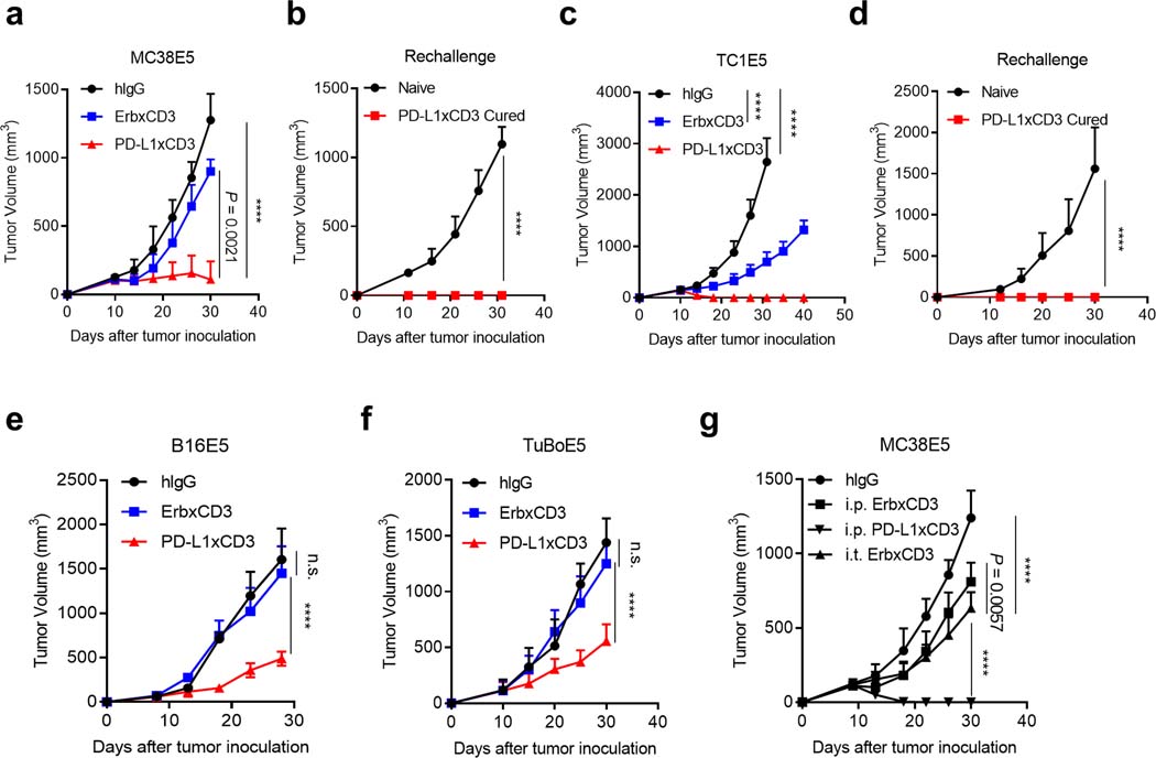
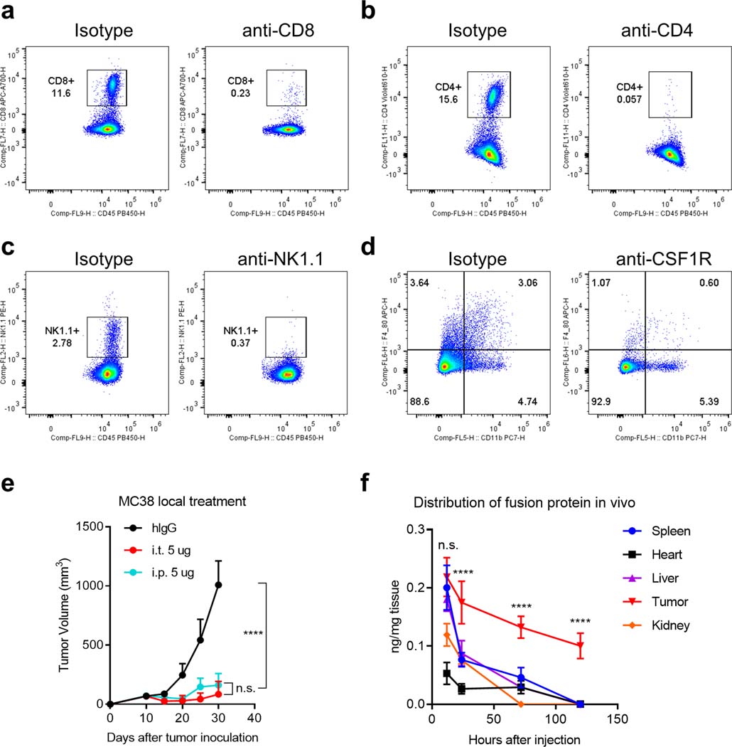
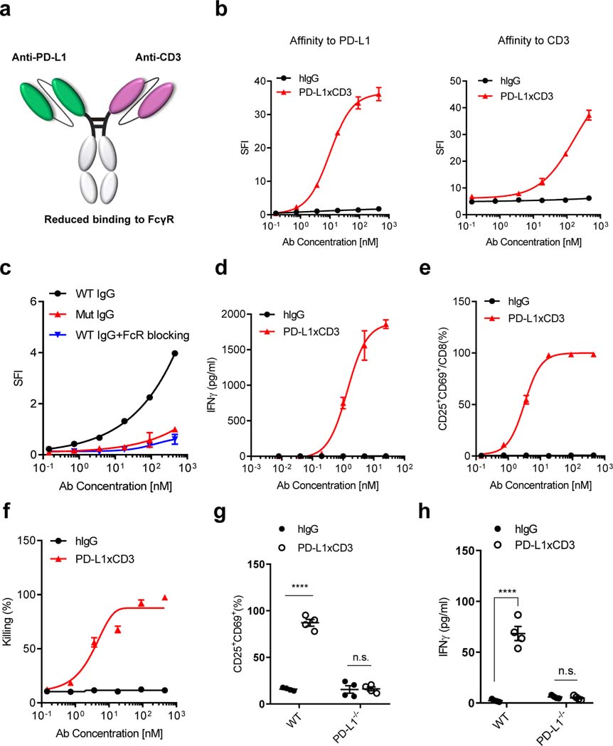
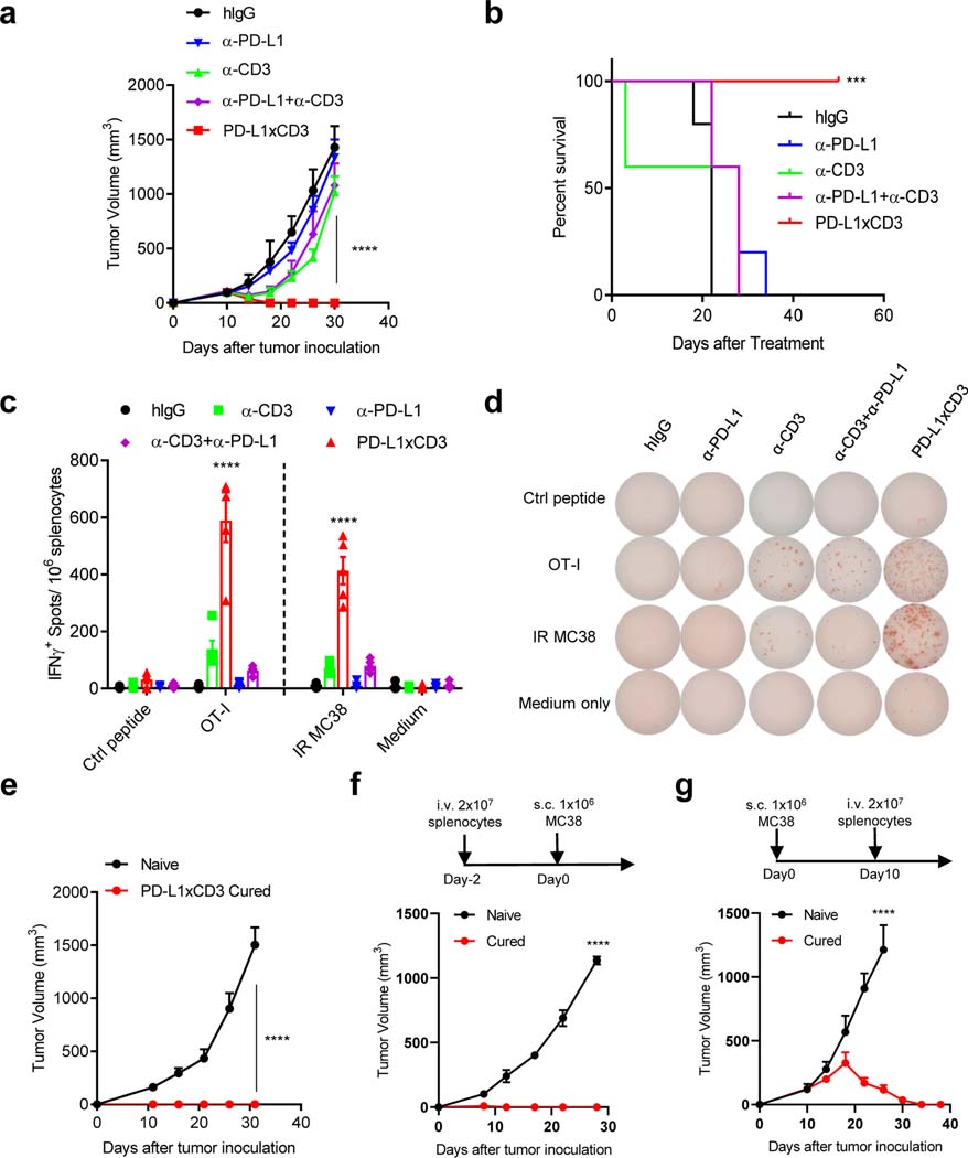
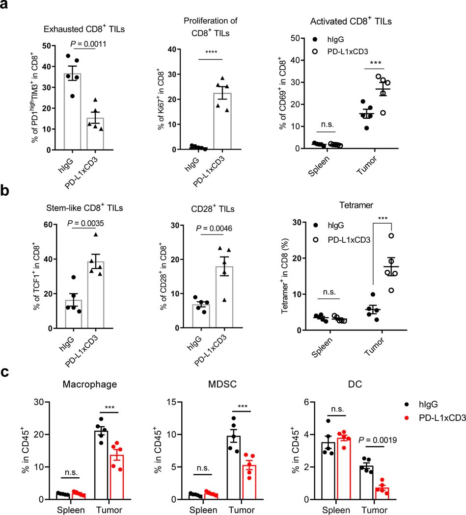
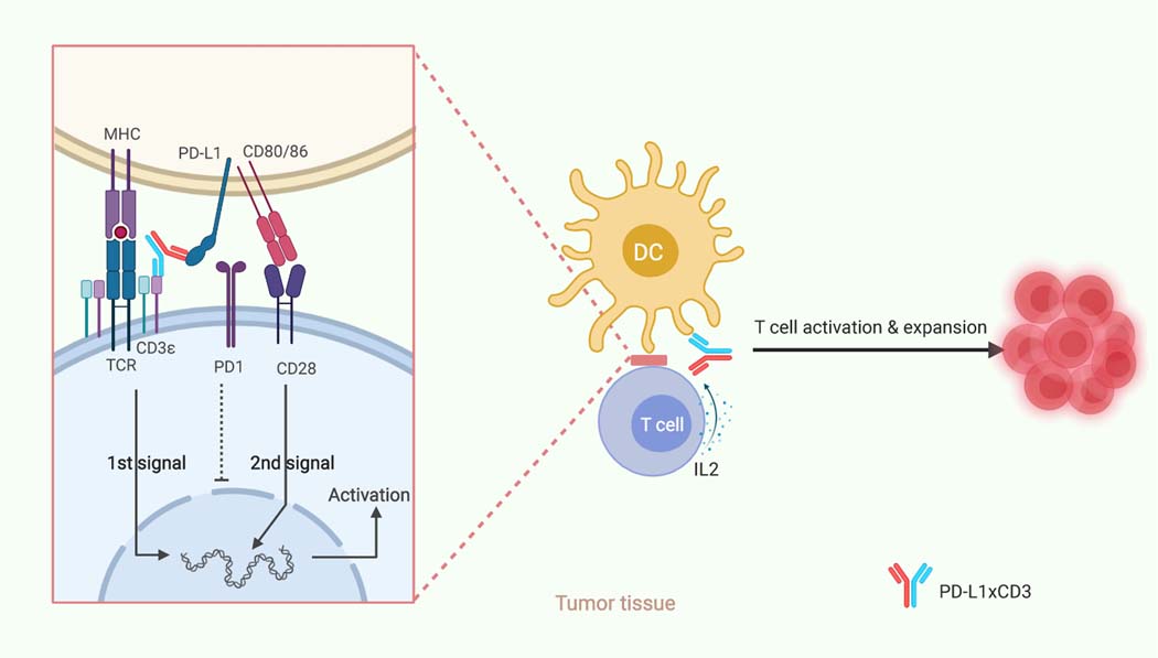
Bispecific T-cell engagers (BiTEs) preferentially targeting tumour-associated antigens and stimulating CD3-mediated signalling are being used in patients to treat acute B-cell lymphoblastic leukemia. However, the potency of BiTEs in solid tumours is limited by their short half-life and their severe toxicity at relevant therapeutic doses. Here we report the design and in vivo performance of a bispecific antibody that simultaneously targets the murine T-cell co-receptor CD3ε and the murine immune checkpoint programmed-death ligand 1 (PD-L1). In multiple syngeneic tumour models, the bispecific antibody generated higher antitumour immune responses than conventional BiTEs targeting tumour-associated antigens and CD3ε. We found that the durable antigen-specific T-cell responses resulted from the rejuvenation of CD8 T cells, owing to the blockade of PD-L1 on dendritic cells (but not on tumour cells) and co-stimulation by B7-1&2 (a peripheral membrane protein on dendritic cells). Bispecific T-cell engagers targeting dendritic cells rather than tumour cells may represent a general means of T-cell rejuvenation for durable cancer immunotherapy.


Conflict of interest statement

Competing interests

The authors declare no competing interests.

Figures

Extended Data Fig. 1 |
Extended Data Fig. 1 |. PD-L1xCD3 generates superior antitumour effects than TAA-targeting BiTE in vivo.
a-b, C57BL/6 J mice were subcutaneously inoculated with 3 × 105 MC38E5 tumor cells and treated with 0.25 mg kg−1 of bispecific antibodies twice on day 10 and 15. Tumor volume was measured twice per week (a). 60 days post treatment, tumor free mice were re-challenged with 3 × 106 tumor cells (b). c-d, C57BL/6 J mice were subcutaneously inoculated with 1 × 106 TC1E5 tumor cells and treated with 0.25 mg kg−1 of bispecific antibodies twice on day 10 and 15. Tumor volume was measured twice per week (c). 60 days post treatment, tumor free mice were re-challenged with 1 × 107 tumor cells (d). e, C57BL/6 J mice were subcutaneously inoculated with 3 × 105 B16E5 tumor cells and treated with 0.25 mg kg−1 of bispecific antibodies intraperitoneally twice on day 8 and 12. f, BALB/c mice were subcutaneously inoculated with 5 × 105 TuBoE5 tumor cells and treated with 0.25 mg kg−1 of fusion proteins intraperitoneally twice on day 10 and 14. g, C57BL/6 J mice were subcutaneously inoculated with 3 × 105 MC38E5 tumor cells and treated with 0.25 mg kg−1 of fusion proteins either intratumorally or intraperitoneally twice on day 9 and 13. Data were presented as mean ± s.e.m from a representative experiment (n = 5 (a, b, f, g), 4 (c-e) biologically independent animals) of two independent experiments. Statistical analysis was performed by two-way ANOVA with Tukey’s multiple comparisons test. ****P ≤ 0.0001.
Extended Data Fig. 2 |
Extended Data Fig. 2 |. PD-L1xCD3 targets pre-existing CD8 T cells in the tumour tissue to initiate the antitumour immune response.
a-d, C57BL/6 J mice were inoculated with 1 × 106 MC38 tumor cells and treated with PD-L1xCD3 (0.25 mg kg−1 on day 10 and 15). 200 μg of anti-CD8, anti-CD4, anti-NK1.1 or 500 μg of anti-CSF1R was administrated respectively one day before treatment initiation and then twice a week for 2 weeks. The percentage of CD8 +cells (a), CD4 +cells (b), NK1.1+ cells (c) and CD11b +F4/80+ cells (d) in the spleen were detected by flow cytometry. e, C57BL/6 J mice (n = 5 biologically independent animals) were subcutaneously inoculated with 1 × 106 MC38 tumor cells and treated with 0.25 mg kg−1 of PD-L1xCD3 either intratumorally or intraperitoneally twice on day 10 and 15. f, C57BL/6 J mice (n = 3 biological replicates) were subcutaneously inoculated with 1 × 106 MC38 tumor cells and intraperitoneally treated with 0.25 mg kg−1 of PD-L1xCD3. Concentration of fusion protein in different tissues were measured by hIgG ELISA at indicated time point. Data were shown as mean ± s.e.m from a representative experiment of two independent experiments. Statistical analysis was performed by two-way ANOVA with Tukey’s multiple comparisons test. ****P ≤ 0.0001.
Extended Data Fig. 3 |
Extended Data Fig. 3 |. Co-stimulatory signaling blockade abolished PD-L1xCD3 mediated antitumour effects and the in vitro activation of T cells by TAA-targeting BiTE.
a-b, C57BL/6 J mice were inoculated with 1 × 106 MC38 tumor cells and treated with PD-L1xCD3 (0.25 mg kg−1 on day 10 and 15), 200 μg CTLA4-Ig was administrated on day 10, 13 and 15. Experimental design (a) and tumor growth curve (b) are shown. c-e, CD8 T cells were co-cultured with either tumor cells or dendritic cells in the presence of ErbxCD3. T cell activation (c), apoptotic T cells (d), supernatant IL-2 and IFN-γ (e) were measured by flow cytometry. Data were shown as mean ± s.e.m from a representative experiment of two independent experiments (n = 5 biologically independent animals). Statistical analyses were performed by two-way ANOVA with Dunnett’s multiple comparisons test (b), two-tailed unpaired Student’s t-test (c-e). ***P ≤ 0.001, ****P ≤ 0.0001.
Extended Data Fig. 4 |
Extended Data Fig. 4 |. Correlation analysis of CD28 expression with CD80/86 expression, dendritic cell infiltration and patient survival.
TCGA database was analyzed for cumulative survival according to CD28 expression (a), correlation of CD28 level with CD80 and CD86 level (b) and correlation of CD28 level with dendritic cell infiltration (c). Skin cutaneous melanoma (SKCM), cervical squamous cell carcinoma and endocervical adenocarcinoma (CESC), lung adenocarcinoma (LUAD), colon adenocarcinoma (COAD), breast invasive carcinoma (BRCA). Statistical analyses were performed by log-rank test (a), and Spearman’s rho correlation test (b-c).
Fig. 1 |
Fig. 1 |. PD-L1xCD3 targets PD-L1 to activate T cells in vitro.
a, Schematic structure of PD-L1xCD3 bispecific antibody. PD-L1xCD3 is composed of a single-chain variable fragment (ScFv) to PD-L1 and a ScFv to murine CD3ε, fused to a mutant human IgG1. b, Binding of PD-L1xCD3 to PD-L1 on MC38 cells overexpressing PD-L1. Cells were incubated with serial dilutions of PD-L1xCD3 or human IgG control, followed by a fluorophore-conjugated anti-human IgG secondary antibody. Flow cytometry measured specific fluorescence index (SFI) using the mean fluorescence intensity (MFI). Binding of PD-L1xCD3 to CD3ε on CD8 T cells purified from mouse spleen. Cells were incubated with serial dilutions of PD-L1xCD3 or human IgG control, followed by a fluorophore-conjugated anti-human IgG secondary antibody. Flow cytometry measured SFI. c, Binding of PD-L1xCD3 to FcγR on RAW 264.7 cells. Cells were incubated with serial dilutions of WT IgG BsAbs, mutant IgG BsAbs, or WT IgG BsAbs with anti- FcγR, followed by a fluorophore-conjugated anti-human IgG secondary antibody. Flow cytometry measured SFI. d-f, MC38-GFP cells (3×10⁴) and purified splenic CD8 T cells (3×10⁵) were co-cultured with serial dilutions of PD-L1xCD3 or human IgG control for 48 hours. IFNγ in the supernatant was detected by cytokine beads array (CBA) (d). CD25 and CD69 expression on T cells were detected by flow cytometry (e). GFP+ 7AAD- tumor cells (viability) were detected by flow cytometry (f). g-h, MC38 tumor cells (WT or PD-L1−/−, 3×10⁴) and purified splenic CD8 T cells (3×10⁵) were co-cultured with PD-L1xCD3 or human IgG control for 48 hours, T cell activation (g) and IFNγ in the supernatant (h) were detected respectively. Data were presented as means ± SEM from a representative experiment (n=3 (b-f), 4 (h-i) biological replicates) of two independent experiments. Data were analyzed using non-linear best fits for (b-f) and two-tailed unpaired Student’s t-test for (g-h). ****P ≤ 0.0001.
Fig. 2 |
Fig. 2 |. PD-L1xCD3 generates superior anti-tumor effect than combination treatment in vivo.
a-b, C57BL/6J mice were subcutaneously inoculated with 1×10⁶ MC38 tumor cells and treated with 0.25 mg/kg of anti-CD3, anti-PD-L1, anti-CD3 plus anti-PD-L1 or PD-L1xCD3 twice on day 10 and 15. Tumor volume (a) and percentage of survival (b) was shown. c-d, C57BL/6J mice were subcutaneously inoculated with 1×10⁶ MC38OVA tumor cells and treated as in panel a. 25 days after treatment, splenocytes from different treatment groups were isolated and stimulated with either OT-I peptide or irradiated MC38 tumor cell (IR MC38). Antigen specific T cells were detected by Elispot assay. e-g, MC38 bearing C57BL/6J mice were treated with PD-L1xCD3 twice on day 10 and day 15 after tumor inoculation. 50 days after treatment, cured mice were re-challenged with 1×10⁷ MC38 tumor cell (e), 2×10⁷ splenocytes from cured mice were adoptively transferred to Rag1−/− mice two days before MC38 tumor cell inoculation (f), 2×10⁷ splenocytes from cured mice were adoptively transferred to MC38 bearing Rag1−/− mice 10 days after tumor inoculation (g). Data were presented as mean ± SEM from a representative experiment (n=5 (a-e), 4 (f-g) biological replicates) of two independent experiments. Statistical analysis was performed by two-way ANOVA with Tukey’s multiple comparisons test (a, c), Sidak’s multiple comparisons test (e-g) or Log-rank (Mantel-Cox) test (b). ***P ≤ 0.001, and ****P ≤ 0.0001.
Fig. 3 |
Fig. 3 |. Pre-existing CD8 T cells are sufficient for the anti-tumor effect of PD-L1xCD3.
a, Rag1−/− mice were inoculated with 1×10⁶ MC38 tumor cells and treated with PD-L1xCD3 (0.25 mg/kg on day 10 and 15). b, C57BL/6 mice were inoculated with 1×10⁶ MC38 tumor cells and treated with PD-L1xCD3 (0.25 mg/kg on day 10 and 15). 200μg anti-CD8 or anti-CD4 was administrated one day before treatment initiation and then twice a week for 2 weeks. c, C57BL/6 mice were inoculated with 1×10⁶ MC38 tumor cells and treated with PD-L1xCD3 (0.25 mg/kg on day 14 and 18). 20μg FTY720 was administrated one day before treatment initiation and then 10μg every other day for 2 weeks. d, C57BL/6 mice were inoculated with 1×10⁶ MC38 tumor cells and treated with PD-L1xCD3 (0.25 mg/kg on day 10 and 15). 200 μg anti-NK1.1 or 500 μg anti-CSF1R was administrated one day before treatment initiation and then twice a week for 2 weeks. Data were presented as mean ± SEM from a representative experiment (n=5 biologically independent animals) of two independent experiments. Statistical analysis was performed by two-way ANOVA with Sidak’s multiple comparisons test (a) or Dunnett’s multiple comparisons test (b-d). ****P ≤ 0.0001.
Fig. 4 |
Fig. 4 |. PD-L1 on dendritic cells is essential for the anti-tumor effect of PD-L1xCD3.
a, C57BL/6J mice were subcutaneously inoculated with 1×10⁶ MC38-PD-L1−/− tumor cells (a, left) or 5×10⁵ B16-PD-L1−/− tumor cells (a, right) and treated with 0.25 mg/kg of PD-L1xCD3 or human IgG control twice on day 10 and 15 after tumor inoculation. b, PD-L1−/− mice were subcutaneously inoculated with 1×10⁶ MC38 tumor cells and treated with 0.25 mg/kg of PD-L1xCD3 or human IgG control twice on day 10 and 15 after tumor inoculation. c, Zbtb46CrePD-L1f/f mice (c, left) or Lyz2CrePD-L1f/f mice (c, right) were subcutaneously inoculated with 1×10⁶ MC38 tumor cells and treated with 0.25 mg/kg of PD-L1xCD3 or human IgG control twice on day 10 and 15 after tumor inoculation. d, Batf3−/− mice were subcutaneously inoculated with 1×10⁶ MC38 tumor cells and treated with PD-L1xCD3 or human IgG control (0.25 mg/kg on day 10 and 15). Data were presented as mean ± SEM from a representative experiment (n=5 biologically independent animals) of two independent experiments. Statistical analysis was performed by two-way ANOVA with Sidak’s multiple comparisons test. ****P ≤ 0.0001.
Fig. 5 |
Fig. 5 |. PD-L1xCD3 reshapes a distinct immunophenotypic signature in tumor-bearing mice.
a, C57BL/6J mice were subcutaneously inoculated with 1×10⁶ MC38-OVA tumor cells and treated with 0.25 mg/kg of PD-L1xCD3 or human IgG control. Flow cytometry analysis was performed with splenocytes and dissociated tumor samples for the percentage of PD-1high TIM-3+ CD8 T cells (a, left), Ki-67+ CD8 T cells (a, middle), CD69+CD8+ T cells (a, right). b, C57BL/6J mice were subcutaneously inoculated with 1×10⁶ MC38-OVA tumor cells and treated with 0.25 mg/kg of PD-L1xCD3 or human IgG control. Flow cytometry analysis was performed with splenocytes and dissociated tumor samples for the percentage of TCF1+ CD8 T cells (b, left), CD28+ CD8 T cells (b, middle), and tetramer+ cells (b, right). c, MC38 bearing mice were treated with PD-L1xCD3 or human IgG. 48 hours after treatment, the percentage of macrophage (c, left), MDSC (c, middle), and dendritic cell (c, right) were detected by flow cytometry with splenocytes and dissociated tumor samples. Representative result from two independent experiments were shown as mean ± SEM (n=5 biological replicates). Statistical analysis was performed by two-tailed unpaired Student’s t-test (a, left and middle; b, left and middle) or two-way ANOVA with Sidak’s multiple comparisons test (a, right; b, right and c). ***P ≤ 0.001, ****P ≤ 0.0001.
Fig. 6 |
Fig. 6 |. Co-stimulatory signaling is required for PD-L1xCD3 mediated anti-tumor effects.
a, C57BL/6J mice were inoculated with 1×10⁶ MC38 tumor cells and treated with PD-L1xCD3 (0.25 mg/kg on day 10 and 15), 200 μg anti-B7–1 and anti-B7–2 were administrated on day 10, 13 and 15. Experimental design (a, left), tumor growth curve (a, middle) and IFNγ-producing antigen specific CD8 T cells (a, right) were shown. b-e, CD8 T cells were co-cultured with either tumor cells or dendritic cells in the presence of bispecific antibodies. T cell activation (b), supernatant IFNγ (c), apoptotic T cells (d) and supernatant IL-2 (e) were measured by flow cytometry and CBA. f-g, Cumulative survival in colorectal adenocarcinoma (f) and liver hepatocellular carcinoma (g) patients according to CD8 infiltration and CD28 level in TCGA database (top 10% vs bottom 10%). Representative result from two independent experiments were shown as mean ± SEM (n=5 biological replicates). Statistical analysis was performed by one-way ANOVA (a, right and b-e), two-way ANOVA (a, middle) with Tukey’s multiple comparisons test and Log-rank test (f-g). ****P ≤ 0.0001.
Fig. 7 |
Fig. 7 |. Schematic of hypothesized working model.
PD-L1xCD3 will predominantly distribute and accumulate in the tumor tissue, targeting PD-L1 positive cells to reactivate the T cells in close proximity with them. As dendritic cells express the highest level of PD-L1 in the TME, PD-L1xCD3 mainly targets dendritic cells to not only crosslink the CD3 complex for the 1st signal activation but also interrupt PD1-PD-L1 mediated inhibition. Meanwhile, PD-L1xCD3 mediated binding to PD-L1 on DCs will release CD80 from the PD-L1-CD80 heterodimer, which can then bind to CD28 on T cells to provide the 2nd signal for T cell activation. With both the 1st and 2nd signals activated, tumor reactivate T cells will overcome activation induced cell death (AICD) and be expanded via IL-2. Thus, T cells in the tumor tissue will be reactivated for effective anti-tumor immunity and memory T cell differentiation. This schematic was created with BioRender.

Similar articles

Cited by

References

    1. Staerz UD, Kanagawa O & Bevan MJ Hybrid antibodies can target sites for attack by T cells. Nature 314, 628–631 (1985). - PubMed
    1. Garber K Bispecific antibodies rise again. Nature reviews. Drug discovery 13, 799–801 (2014). - PubMed
    1. Baeuerle PA & Reinhardt C Bispecific T-cell engaging antibodies for cancer therapy. Cancer research 69, 4941–4944 (2009). - PubMed
    1. Trabolsi A, Arumov A & Schatz JH T Cell-Activating Bispecific Antibodies in Cancer Therapy. Journal of immunology 203, 585–592 (2019). - PubMed
    1. Bargou R et al. Tumor regression in cancer patients by very low doses of a T cell-engaging antibody. Science 321, 974–977 (2008). - PubMed

Publication types