Review
. 2021 Nov 2;23(23 Suppl 5):S16-S29.
doi: 10.1093/neuonc/noab143.

DNA methylation profiling as a model for discovery and precision diagnostics in neuro-oncology

Affiliations
Review

DNA methylation profiling as a model for discovery and precision diagnostics in neuro-oncology

Drew Pratt et al. Neuro Oncol. .

Abstract

Recent years have witnessed a shift to more objective and biologically-driven methods for central nervous system (CNS) tumor classification. The 2016 world health organization (WHO) classification update ("blue book") introduced molecular diagnostic criteria into the definitions of specific entities as a response to the plethora of evidence that key molecular alterations define distinct tumor types and are clinically meaningful. While in the past such diagnostic alterations included specific mutations, copy number changes, or gene fusions, the emergence of DNA methylation arrays in recent years has similarly resulted in improved diagnostic precision, increased reliability, and has provided an effective framework for the discovery of new tumor types. In many instances, there is an intimate relationship between these mutations/fusions and DNA methylation signatures. The adoption of methylation data into neuro-oncology nosology has been greatly aided by the availability of technology compatible with clinical diagnostics, along with the development of a freely accessible machine learning-based classifier. In this review, we highlight the utility of DNA methylation profiling in CNS tumor classification with a focus on recently described novel and rare tumor types, as well as its contribution to refining existing types.

Keywords: DNA methylation; brain tumor; neuro-oncology.


Figures

Fig. 1
Fig. 1
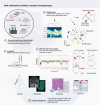
Workflow and applications of DNA methylation profiling in surgical neuropathology. The Illumina 450k and HumanMethylationEPIC (EPIC) arrays are compatible with both fresh/frozen tissue and Formalin-Fixed Paraffin-Embedded (FFPE) material (A). Following extraction, DNA is bisulfite-converted and hybridized to the BeadChip. The fluorescent signals are then processed with the iScan or NextSeq 550 readers and two separate data files (*.idat) are produced, one for each color channel (i.e. red and green). Following various preprocessing steps, methylation data (often represented as beta values) can then be used as input for clustering or data visualization (B). In addition to hierarchical clustering, dimensionality reduction with t-Distributed Stochastic Neighbor Embedding (t-SNE) or Uniform Manifold Approximation and Projection (UMAP) provides a novel technique for confirming and identifying new tumor types. The basis for this method of tumor diagnostics relies on epigenetically (and often genetically) similar tumor types grouping together in the two- or three-dimensional space; subtypes can also be identified within these macro-clusters. Machine learning algorithms, such as random forest, can be used to train a classifier based on pre-defined cluster labels using these dimensionality reduction techniques. The combined intensities of the signals from the methylated and unmethylated channels may be used to infer focal and broad copy number changes (C). Diagnostic markers in central nervous system (CNS) tumors, such as whole-arm 1p/19q codeletion and the +7/−10 signature (with or without EGFR amplification) can be reliably detected with this method; furthermore, copy number breakpoints are useful to infer fusion events in the correct diagnostic context. A two-CpG methylation signature can be used to assess O-6-methylguanine-DNA methyltransferase (MGMT) promoter methylation status and has been independently associated with response to alkylating therapy in isocitrate dehydrogenase (IDH)-wildtype glioblastoma (GBM) (D). Finally, as with any diagnostic assay in surgical pathology, methylation profiling should be interpreted in conjunction with all available clinical data, including history, imaging, histology, and other ancillary molecular techniques such as sequencing (E).
Fig. 2
Fig. 2
Dimensionality reduction techniques for the visual assessment of central nervous system (CNS) tumor clustering. Two frequently used methods for visualizing tumor types are Uniform Manifold Approximation and Projection (UMAP) (A) and t-distributed stochastic neighbor embedding (t-SNE) (B). Illustrated are selected novel and rare CNS tumor types that are discussed in this review (not included: diffuse glioneuronal tumor with oligodendroglioma-like features and nuclear clusters (DGONC) and cribriform neuroepithelial tumor (CRINET), and those presented in “epigenetic subtyping”). As a rule of thumb, the distance between the groups is less meaningful than the relationship within the groups. UMAP is thought to preserve global structure better than t-SNE, but both produce comparable results for classifying CNS tumor types. Abbreviation: AT/RT, atypical teratoid/rhabdoid tumor (TYR, SHH, MYC subtypes); DMT, desmoplastic myxoid tumor; PDC, poorly differentiated chordoma; ETMR, embryonal tumor with multilayered rosettes; BCOR, CNS tumor with BCOR with internal tandem duplication; SARC, DICER1, primary intracranial sarcoma, DICER1-mutant; CIC, CIC-rearranged sarcoma; RGNT, rosette-forming glioneuronal tumor; HGAP, high-grade astrocytoma with piloid features; MYB/L, diffuse astrocytoma, MYB or MYBL1-altered; IHG, infant-type hemispheric glioma; FOXR2, CNS neuroblastoma, FOXR2-activated.
Fig. 3
Fig. 3
Heterogeneity in histologic diagnoses and genetic alterations among central nervous system (CNS) tumor types from Figure 2. Clustering of CNS tumors by methylation profiling frequently reveals remarkable variability in histopathology within tumor types, often with discrepancies in the world health organization (WHO) grade. Additionally, large-scale studies and case reports have also revealed previously unrecognized heterogeneity in the genetic landscape of some tumors. Abbreviation: mut, mutation; fus, fusion.

References

    1. You JS, Jones PA. Cancer genetics and epigenetics: two sides of the same coin? Cancer Cell. 2012;22(1):9–20. - PMC - PubMed
    1. Tost J. DNA methylation: an introduction to the biology and the disease-associated changes of a promising biomarker. Methods Mol Biol. 2009;507:3–20. - PubMed
    1. Jones PA. Functions of DNA methylation: islands, start sites, gene bodies and beyond. Nat Rev Genet. 2012;13(7):484–492. - PubMed
    1. Klughammer J, Kiesel B, Roetzer T, et al. . The DNA methylation landscape of glioblastoma disease progression shows extensive heterogeneity in time and space. Nat Med. 2018;24(10):1611–1624. - PMC - PubMed
    1. Louis DN, Perry A, Reifenberger G, et al. . The 2016 world health organization classification of tumors of the central nervous system: a summary. Acta Neuropathol. 2016;131(6):803–820. - PubMed

Publication types