. 2021 Nov 1;77(Pt 11):388-398.
doi: 10.1107/S2053230X2100964X. Epub 2021 Oct 19.

The crystal structures of the enzyme hydroxymethylbilane synthase, also known as porphobilinogen deaminase

Affiliations

The crystal structures of the enzyme hydroxymethylbilane synthase, also known as porphobilinogen deaminase

John R Helliwell. Acta Crystallogr F Struct Biol Commun. .

Abstract

The enzyme hydroxymethylbilane synthase (HMBS; EC 4.3.1.8), also known as porphobilinogen deaminase, catalyses the stepwise addition of four molecules of porphobilinogen to form the linear tetrapyrrole 1-hydroxymethylbilane. Thirty years of crystal structures are surveyed in this topical review. These crystal structures aim at the elucidation of the structural basis of the complex reaction mechanism involving the formation of tetrapyrrole from individual porphobilinogen units. The consistency between the various structures is assessed. This includes an evaluation of the precision of each molecular model and what was not modelled. A survey is also made of the crystallization conditions used in the context of the operational pH of the enzyme. The combination of 3D structural techniques, seeking accuracy, has also been a feature of this research effort. Thus, SAXS, NMR and computational molecular dynamics have also been applied. The general framework is also a considerable chemistry research effort to understand the function of the enzyme and its medical pathologies in acute intermittent porphyria (AIP). Mutational studies and their impact on the catalytic reaction provide insight into the basis of AIP and are also invaluable for guiding the understanding of the crystal structure results. Future directions for research on HMBS are described, including the need to determine the protonation states of key amino-acid residues identified as being catalytically important. The question remains - what is the molecular engine for this complex reaction? Thermal fluctuations are the only suggestion thus far.

Keywords: enzyme–substrate intermediates; hydroxymethylbilane synthase; porphobilinogen deaminase; reaction mechanisms; structure and function.


Figures

Figure 1
Figure 1
The biosynthesis of uroporphyrinogen III from 5-aminolevulinic acid. The enzymes involved are (a) 5-aminolaevulinic acid dehydratase, (b) hydroxymethylbilane synthase (HMBS) and (c) uroporphyrinogen III synthase. A = CH2COO; P = CH2CH2COO. Reproduced from Hädener et al. (1999 ▸).
Figure 2
Figure 2
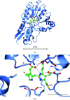
(a) The crystal structure of the active form of the E. coli HMBS enzyme in ribbon format (PDB entry 1ah5); the cofactor is in the middle of the picture. (b) An enlargement of the dipyrromethane cofactor; the right-hand cofactor ring (referred to in the text as C1) is covalently attached to the labelled Cys242 (Cys261 in the human enzyme). The Asp side chain (Asp84 in E. coli HMBS and Asp99 in human HMBS) is visible just below the cofactor towards its left-hand side. This figure was produced by CCP4mg (McNicholas et al., 2011 ▸).
Figure 3
Figure 3
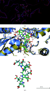
Best least-squares-calculated overlay of the cofactor plus ES2 for human HMBS, PDB entries 5m6r and human 7aak, in molecule A. Molecule B also shows a similarly good agreement between PDB entries 5m6r and 7aak. (a) was made with Coot (Emsley et al., 2010 ▸), and (b) and (c) were made with CCP4mg (McNicholas et al., 2011 ▸). All three show very similar orientations and show complementary information. Cys261 is covalently linked to the first ring of the cofactor. The least-squares-calculated overlay of PDB entries 5m6r and 7aak used the five amino acids centred on Asp99.
Figure 4
Figure 4
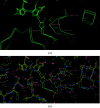
The motion of (human) Cys261 itself is largely responsible for pulling the cofactor to make room for the addition of two PBG molecules to form ES2 (Bustad et al., 2021 ▸). The movement of Cys261 (PDB entries 7aaj and 7aak, molecules A) is 4.4 Å and that of Val263 is 0.8 Å. The movement of Asp99 is 0.5 Å. (a) shows the alpha carbons and the cofactors and ES2; (b) shows an identical view with all atoms.
Figure 5
Figure 5
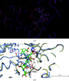
Best least-squares-calculated overlay of the cofactor plus ES2 for human HMBS, PDB entries 5m6r and human 7cd0, in molecule A. Molecule B shows very similar agreement. Cys261 is at the lower middle and thereby also identifies the first ring of the cofactor. Note that the iodinated PBG inhibitor is in molecule B. (a) was made with Coot (Emsley et al., 2010 ▸) and (b) was made with CCP4mg (McNicholas et al., 2011 ▸). Both show very similar orientations and show complementary information. Cys261 is covalently linked to the first ring of the cofactor. The least-squares-calculated overlay of PDB entries 5m6r and 7cd0 used the five amino acids centred on Asp99.
Figure 6
Figure 6
Crystal-packing diagram for E. coli (PDB entry 1ypn) showing the solvent channel directly above the 242–255 polypeptide loop of HMBS. Note that the lattice neighbour of Gly255 is Gly33 and residue 32 is a proline, i.e. it is unlikely to interfere with loop movement.
Figure 7
Figure 7
Stereoview of the modelling of the Michaelis complex EP (red) overlaid with the 2 h time point (F oF c) electron-density omit map contoured at 2.00σ. From Nieh (1997 ▸). The acetate and propionate side chains of the second, third and fourth pyrroles could not be placed due to insufficient detail in the density map.

References

    1. Azim, N., Deery, E., Warren, M. J., Erskine, P., Cooper, J. B., Wood, S. P. & Akhtar, M. (2013). Acta Cryst. F69, 906–908. - PMC - PubMed
    1. Azim, N., Deery, E., Warren, M. J., Wolfenden, B. A. A., Erskine, P., Cooper, J. B., Coker, A., Wood, S. P. & Akhtar, M. (2014). Acta Cryst. D70, 744–751. - PMC - PubMed
    1. Bung, N., Pradhan, M., Srinivasan, H. & Bulusu, G. (2014). PLoS Comput. Biol. 10, e1003484. - PMC - PubMed
    1. Bung, N., Roy, A., Chen, B., Das, D., Pradhan, M., Yasuda, M., New, M. I., Desnick, R. J. & Bulusu, G. (2018). Proc. Natl Acad. Sci. USA, 115, E4071–E4080. - PMC - PubMed
    1. Bung, N., Roy, A., Priyakumar, U. D. & Bulusu, G. (2019). Phys. Chem. Chem. Phys. 21, 7932–7940. - PubMed

MeSH terms

Substances